Bone
Bones are rigid organs forming the vertebrate skeleton.

Donvikro · CC BY-SA 4.0
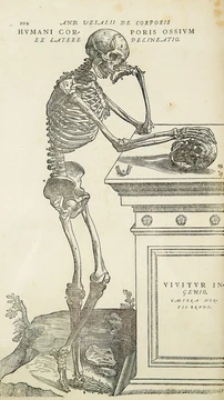
Bones are the rigid organs that make up the skeleton of most vertebrate animals. They shield internal organs, generate red and white blood cells, hold mineral reserves, assist in maintaining acid-base balance, give the body its structure and support, and make movement and hearing possible. Bone tissue—also called osseous tissue—is a hardened, specialized connective tissue that becomes mineralized and features a honeycomb-like intercellular matrix, which gives bone its stiffness. This tissue contains several types of cells: osteoblasts and osteocytes (which build and mineralize bone, respectively), osteoclasts (which break down bone), and flattened or modified osteoblasts known as lining cells, which form a protective layer on the bone’s surface. The mineralized matrix has an organic part made mostly of ossein (a form of collagen) and an inorganic part composed of various salts known as bone mineral. Bone tissue comes in two main forms: cortical (compact) bone and cancellous (spongy) bone, though bones may also contain marrow, endosteum, periosteum, nerves, blood vessels, and cartilage.
At birth, the human body has about 300 bones. Many of these fuse as a person grows, leaving 206 separate bones in an adult, not counting the many small sesamoid bones. The femur, or thighbone, is the largest bone, while the stapes in the middle ear is the smallest. The Ancient Greek word for bone is ὀστέον ("osteon"). In anatomical terminology, including the Terminologia Anatomica, the word for a bone is *os* (for example, *os breve*, *os longum*, *os sesamoideum*). This should not be confused with the alternative medical use of *os* to mean an opening, which comes from the Latin *ōs* for mouth.
**Gross anatomy** Five types of bones exist in the human body: long, short, flat, irregular, and sesamoid. Long bones have a shaft (the diaphysis) that is much longer than it is wide, and a rounded head (the epiphysis) at each end. They consist mostly of compact bone, with some marrow inside the medullary cavity and areas of spongy, cancellous bone at the ends. Most limb bones, including those in the fingers and toes, are long bones, except for the eight carpal bones of the wrist, the seven articulating tarsal bones of the ankle, and the kneecap sesamoid. Long bones with differently shaped shafts or ends, like the clavicle, are called modified long bones. Short bones are roughly cube-shaped, with a thin layer of compact bone around a spongy interior; they offer stability, support, and limited motion. The wrist and ankle bones are short bones. Flat bones are thin and usually curved, with two parallel layers of compact bone sandwiching a layer of spongy bone; most skull bones and the sternum are flat bones. Sesamoid bones are embedded in tendons; by holding the tendon farther from the joint, they increase the tendon’s angle and thus the muscle’s leverage. Examples include the patella and the pisiform. Irregular bones do not fit the other categories; they have thin layers of compact bone around a spongy interior and often have complicated shapes due to multiple ossification centers or bony sinuses. The spine, pelvis, and some skull bones (like the ethmoid and sphenoid) are irregular bones.
**Terminology** Anatomists use various terms from Latin and Greek to describe bones’ appearance, shape, and function. Some still use Latin for bone names. The words “osseous” and the prefix “osteo-” are still common. For example, a “foramen” is a hole through which something passes, while a “canal” or “meatus” is a tunnel. Protrusions from a bone are called a “condyle,” “crest,” “spine,” “eminence,” “tubercle,” or “tuberosity,” depending on shape and location. Long bones are said to have a “head,” “neck,” and “body.” When two bones join, they “articulate.” If the connection is fibrous and relatively immobile, the joint is called a “suture.”
**Functions** *Mechanical*: Bones together form the skeleton, providing a frame that supports the body and serves as attachment points for muscles, tendons, ligaments, and joints. These structures work together to generate and transfer forces, allowing body parts or the whole body to move in three-dimensional space (studied in biomechanics). Bones also protect internal organs—for instance, the skull shields the brain, and the ribs protect the heart and lungs. Bone has a high compressive strength (about 170 MPa), a lower tensile strength (104–121 MPa), and a very low shear strength (51.6 MPa). This means it resists pushing forces well, resists pulling forces less well, and handles twisting or shear forces poorly. Although bone is brittle, it has significant elasticity, mainly due to collagen. Mechanically, bones also play a special role in hearing: the three small ossicles in the middle ear are involved in sound transduction.
*Synthetic*: The cancellous portion of bone is involved in metabolic functions such as mineral storage and blood cell production.
- largest_bone
- femur (thigh-bone)
- smallest_bone
- stapes (in the middle ear)
- ancient_greek_term
- ὀστέον (osteon)
- anatomical_term
- os
Lore & Background
Bones come in a variety of shapes and sizes with complex internal and external structures. The largest bone is the femur, and the smallest is the stapes in the middle ear. Five types of bones are found in the human body: long, short, flat, irregular, and sesamoid. Long bones, such as those of the limbs, have a shaft (diaphysis) and rounded ends (epiphyses). Short bones are roughly cube-shaped, flat bones are thin and curved, sesamoid bones are embedded in tendons, and irregular bones have complicated shapes.
Reader's Guide
Bones serve mechanical, synthetic, and metabolic functions. Mechanically, they form the skeleton, provide support, protect organs, enable hearing via the ossicles, and have high compressive strength but lower tensile and shear strength. Synthetically, bone marrow produces blood cells through hematopoiesis, including red blood cells, platelets, and white blood cells. Metabolically, bones store minerals such as calcium and phosphorus, store growth factors and fatty acids, buffer blood pH, store heavy metals for detoxification, and act as an endocrine organ by releasing fibroblast growth factor 23 and osteocalcin to regulate phosphate metabolism and blood sugar. Bone tissue is continuously remodeled by specialized cells and consists of a flexible matrix (about 30%) and bound minerals (about 70%), making it hard and strong yet lightweight.
Did You Know?
- The largest bone in the body is the femur, and the smallest is the stapes in the middle ear.
- Bone marrow produces over 2.5 billion red blood cells and platelets daily.
- Bones act as an endocrine organ, releasing FGF-23 and osteocalcin to regulate phosphate and glucose.
Anatomy & Structure in the Human Foot
The navicular bone occupies a distinctive position among the tarsal bones of the human foot, sitting on the medial side and serving as a connector between several neighboring structures. Its name comes from its visual resemblance to a tiny boat, a shape created by the deeply concave surface on its proximal, or posterior, side. Anatomically, it forms joints with the talus above it, the three cuneiform bones below, and the cuboid on its lateral side. In terms of development, the navicular is notably the last of the foot's bones to begin the ossification process, typically not starting until around the fourth year of life, though considerable individual variation has been documented. Only a single muscle, the tibialis posterior, attaches directly to this bone, with its main portion inserting into the navicular's tuberosity. Additionally, a small percentage of the general population—estimated between two and fourteen percent—possess an accessory navicular bone, a variant that adds further complexity to this already intricate region of the foot.
Biomechanical Keystone & Injury Patterns
Often described as the keystone of the foot, the navicular bone plays a central structural and functional role in human gait. As a component of the coxa pedis, it articulates with the talus, all three cuneiform bones, the cuboid, and the calcaneus, making it a critical structural link bridging the midfoot and forefoot. This positioning allows the navicular to contribute meaningfully to inversion, eversion, and overall foot motion, while also forming part of both the longitudinal and transverse arches that define the foot's characteristic shape. Despite its importance, the navicular is not a frequently fractured bone. When it does break, the mechanism is typically one of two: a stress fracture, which occurs commonly among athletes subjected to repetitive loading, or a high-energy traumatic event. The relative rarity of navicular fractures, combined with the bone's deep anatomical position and its multiple articulations, makes identification and management of these injuries a particular challenge for clinicians.
The Equine Navicular: A Different Bone with the Same Name
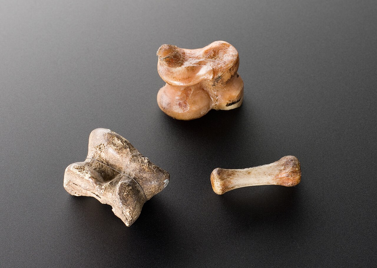
In horses, the term navicular bone refers to an entirely different anatomical structure than the one found in the human foot. The equine navicular is a sesamoid bone situated within the hoof, resting on the palmar aspect of the coffin joint between the second phalanx and the third phalanx, also known as the coffin bone. It is held in place by the distal sesamoidean impar ligament along with two collateral sesamoidean ligaments. A fluid-filled sac called the navicular bursa sits between the flexor surface of this bone and the deep digital flexor tendon, which itself runs between the bursa and the distal phalanx. While the central tarsal bone in the horse's hock is considered homologous and analogous to the human foot's navicular, the sesamoid navicular in the hoof is a fundamentally distinct structure. This naming overlap between species can create considerable confusion in comparative anatomy discussions, as the two bones share a name yet differ in location, shape, and function.
Navicular Disease & the Diagnostic Challenge in Horses
The navicular region in horses carries enormous clinical weight, particularly in the front feet, where it is implicated in a significant and often frustrating condition known as navicular disease or navicular syndrome. This condition may be responsible for as much as one-third of all lameness cases in horses, making it one of the most prevalent causes of impaired mobility in the species. However, diagnosing navicular syndrome has proven far more complex than earlier literature suggested. Much of the original body of research, particularly the reliance on radiographic changes in the navicular bone as a sole diagnostic criterion, has been called into question. Radiographic findings do not always yield a definitive diagnosis, and newer imaging techniques have revealed that damage to the surrounding soft tissues in the navicular region may be a significant contributor to lameness. Furthermore, multiple distinct causes can produce visible lameness, meaning that the navicular bone's appearance on an X-ray is only one piece of a much larger diagnostic puzzle.
Gallery
Frequently Asked Questions
What is a bone?
A bone is a rigid structural organ that forms part of the skeleton in most vertebrate animals. It acts as both a protective framework and a functional unit within the body.
What are a bone's main functions?
Bones shield internal organs, generate red and white blood cells, and store essential minerals. They also help regulate the body's acid-base balance, provide structural support, and enable movement and hearing.
What is the largest bone and what is the smallest?
The femur, or thigh bone, holds the title of the largest bone in the vertebrate body. The smallest is the stapes, a tiny structure located in the middle ear.
What is bone tissue made of?
Bone tissue, also called osseous tissue, is a mineralized form of hard connective tissue with a honeycomb-like matrix. It contains several specialized cell types, including osteoblasts, osteocytes, osteoclasts, and lining cells.
Where does the term 'bone' come from?
The anatomical term for bone is 'os,' while the ancient Greek word for it is osteon (ὀστέον). These roots appear in many related medical and biological terms.
More in Animal Anatomy 1-17
Spotted an error? Know more?
This is a living reference — every entry is fact-audited, and reader corrections feed straight into our audit queue. Suggest an edit · See this site's audit record