Cartilage
Resilient connective tissue covering joints and supporting body structures.

Cristobal carrasco (originale); Fabyrav · CC BY-SA 3.0
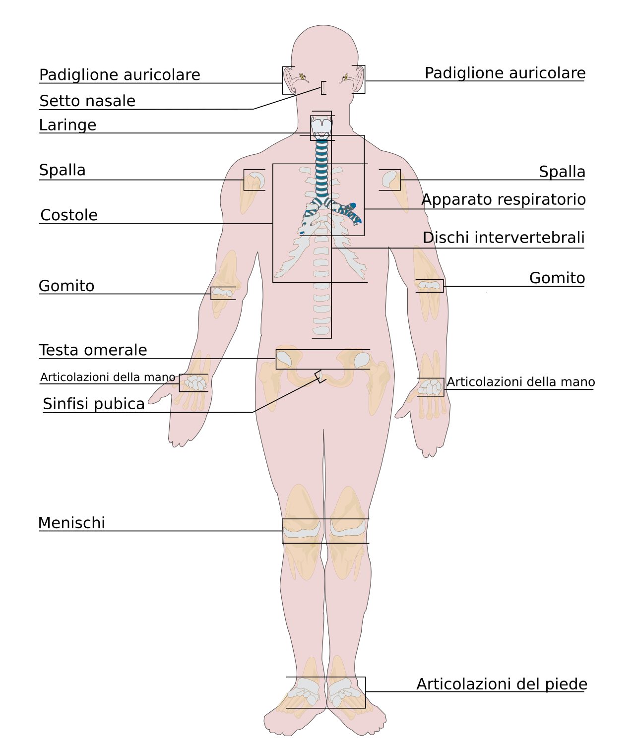
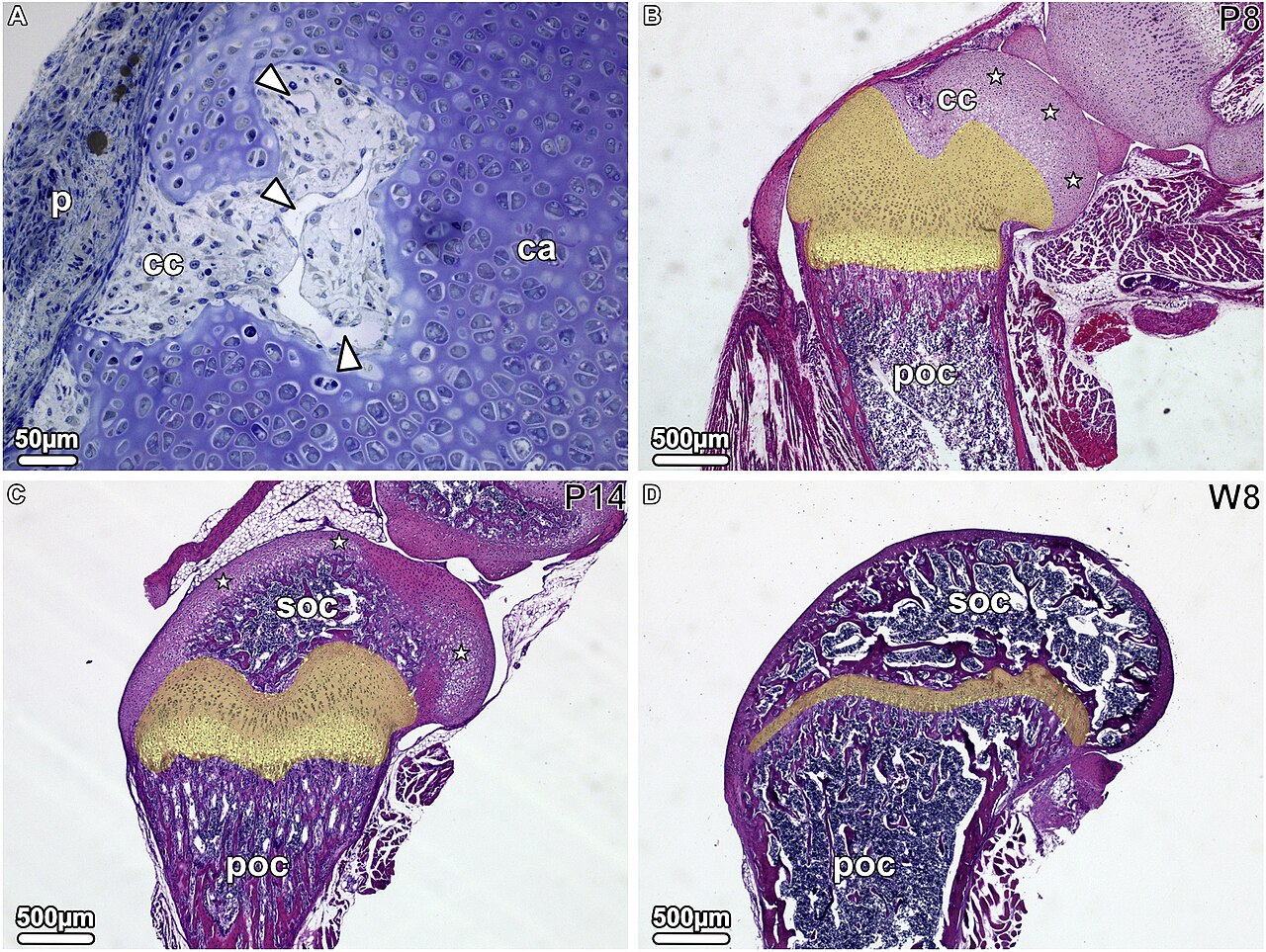
Cartilage is a smooth, resilient connective tissue that is semi-transparent and non-porous, typically encased in a tough, fibrous membrane known as the perichondrium. In tetrapods, it forms articular cartilage, which cushions the ends of long bones at joints, and also appears in structures like the rib cage, neck, bronchial tubes, and intervertebral discs. In other groups, such as chondrichthyans and cyclostomes, cartilage makes up a much larger portion of the skeleton. While not as hard as bone, it is significantly stiffer and less flexible than muscle or tendon. Its rigidity helps keep body tubes open, as seen in the tracheal rings, including the cricoid cartilage and carina.
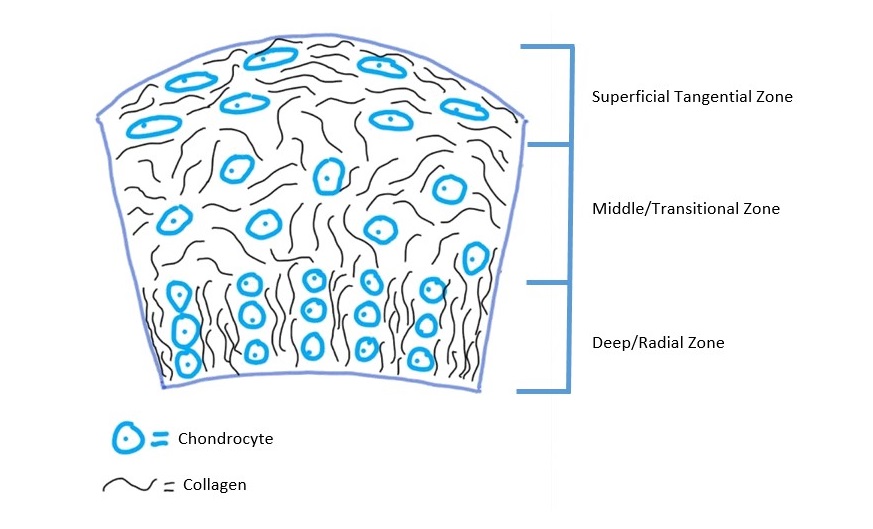
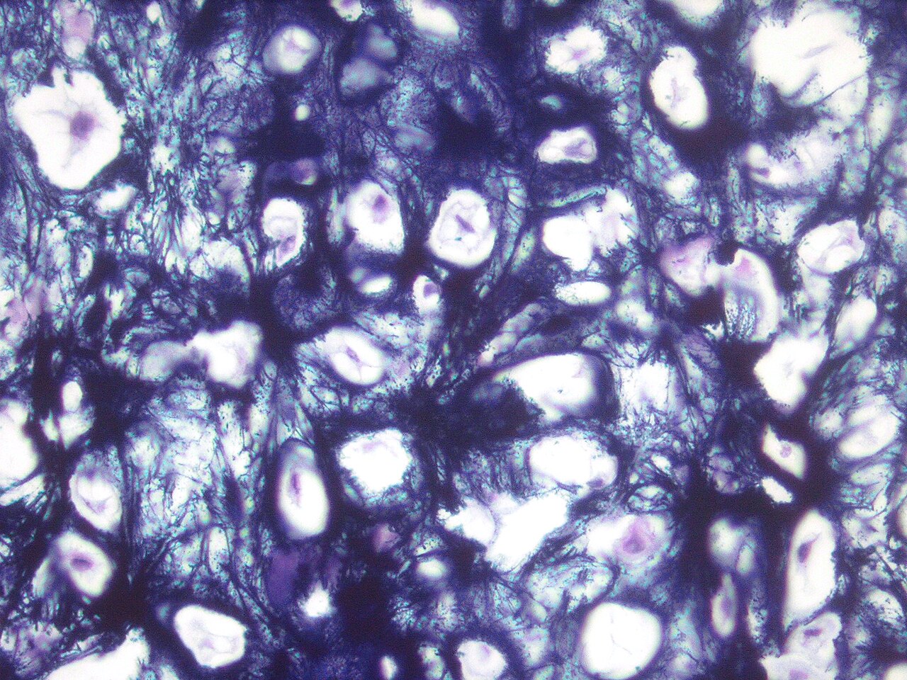
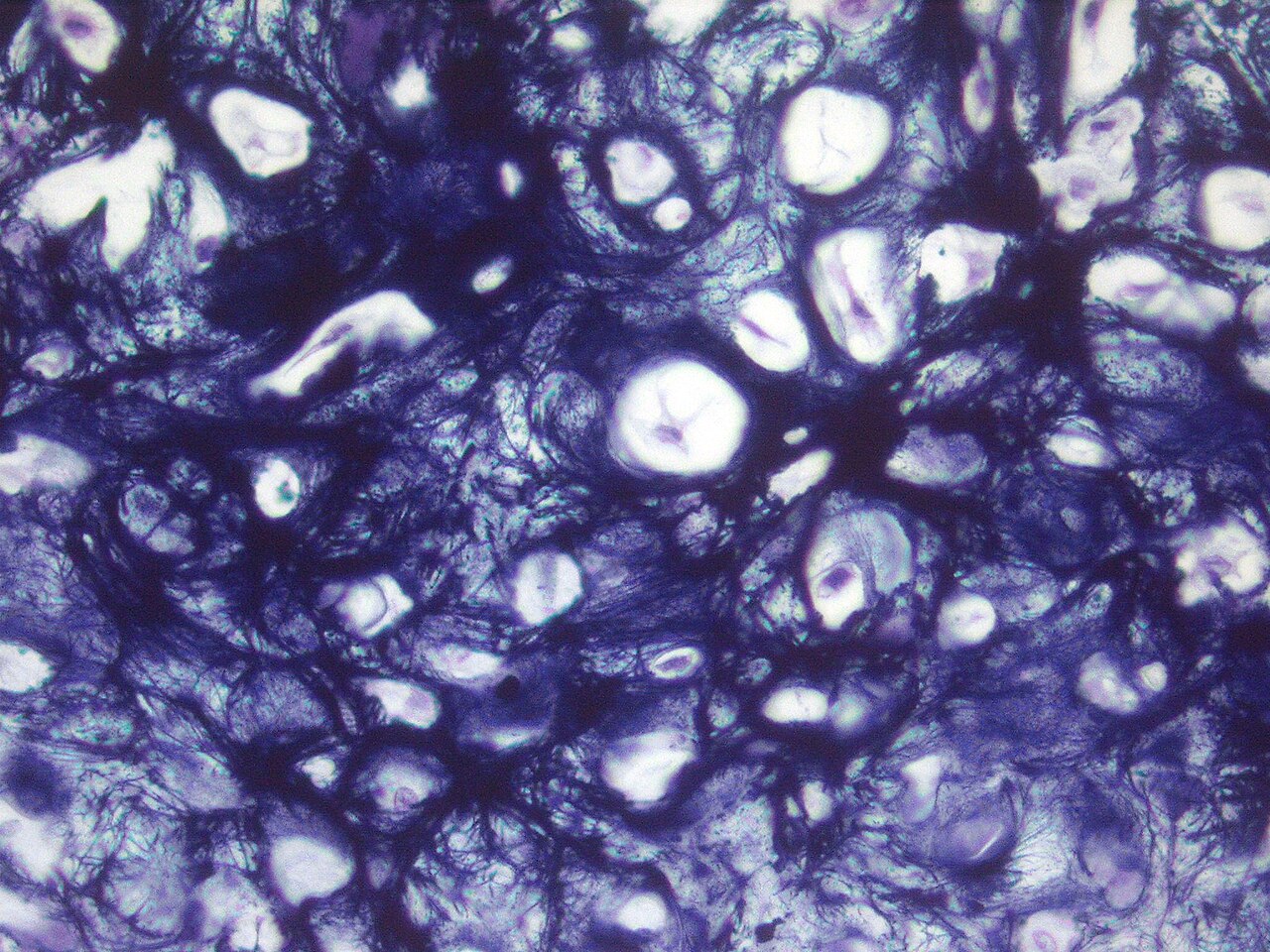
Cartilage consists of specialized cells called chondrocytes, which produce a collagen-rich extracellular matrix with abundant ground substance containing proteoglycans and elastin fibers. The matrix itself is composed of glycosaminoglycans, proteoglycans, collagen fibers, and sometimes elastin. Cartilage grows faster than bone and is classified into three types—elastic cartilage, hyaline cartilage, and fibrocartilage—based on their varying proportions of collagen and proteoglycan. Lacking blood vessels and nerves, cartilage is generally insensitive, though some fibrocartilage, like the knee meniscus, has a partial blood supply. Chondrocytes receive nutrients via diffusion, aided by fluid flow generated when articular cartilage is compressed or elastic cartilage is flexed. Compared to other connective tissues, cartilage has a very slow matrix turnover and repairs only at a very slow rate.
During embryogenesis, the skeletal system arises from the mesoderm germ layer. Cartilage forms through chondrification, where condensed mesenchyme tissue differentiates into chondroblasts that secrete molecules like aggrecan and collagen type II to build the extracellular matrix. In all vertebrates, cartilage is the primary skeletal tissue early in development; in osteichthyans, many cartilaginous elements later ossify via endochondral and perichondral ossification. After initial chondrification, cartilage growth mainly involves maturing immature cartilage, as cell division within cartilage is very slow and growth typically does not come from increasing size or mass. Non-coding RNAs, such as miRNAs and long non-coding RNAs, act as key epigenetic modulators that can influence chondrogenesis and are involved in cartilage-related conditions like arthritis.
The function of articular cartilage depends on the molecular makeup of its extracellular matrix, which is primarily composed of proteoglycans and collagens. The main proteoglycan, aggrecan, forms large aggregates with hyaluronan and itself; these aggregates are negatively charged and retain water in the tissue. Collagen, mostly type II, constrains the proteoglycans, and the matrix responds to tensile and compressive forces. Cartilage growth refers to matrix deposition, as well as the growth and remodeling of the extracellular matrix. Due to high stress on the patellofemoral joint during resisted knee extension, the articular cartilage of the patella is among the thickest in the human body. The ECM of articular cartilage is divided into three regions: pericellular, territorial, and interterritorial matrix.
The mechanical properties of articular cartilage in load-bearing joints like the knee and hip have been studied at macro, micro, and nano-scales, including its responses to frictional, compressive, shear, and tensile loading. Cartilage is resilient and exhibits viscoelastic properties. Because it contains free-moving interstitial fluid, testing is challenging. A common method is the confined compression test, used in creep mode (measuring displacement over time under constant load) or relaxation mode (measuring force over time under constant displacement). In this test, deformation occurs in two main regions: first, rapid displacement from fluid outflow, then slower displacement until a constant equilibrium is reached, which can take hours under typical conditions. In both modes, a disc of cartilage is placed in an impervious, fluid-filled container and covered with a porous plate that restricts fluid flow to the vertical direction. This test measures the aggregate modulus (typically 0.5 to 0.9 MPa for articular cartilage), which indicates tissue stiffness at equilibrium when fluid flow has stopped, and Young's modulus (typically 0.45 to 0.80 MPa), which measures how much a material strains under a given stress.
- composition
- Matrix of glycosaminoglycans, proteoglycans, collagen fibers, and sometimes elastin
- cell type
- Chondrocytes
- types
- Elastic cartilage, hyaline cartilage, and fibrocartilage
- blood supply
- Does not contain blood vessels or nerves; some fibrocartilage has partial blood supply
- mechanical properties
- Viscoelastic; aggregate modulus 0.5–0.9 MPa; Young's modulus 0.45–0.80 MPa
- permeability
- 10^-15 to 10^-16 m^4/Ns
- Poisson's ratio
- Around 0.4 or lower in humans; 0.46–0.5 in bovine subjects
Lore & Background
Cartilage is composed of specialized cells called chondrocytes that produce a large amount of collagenous extracellular matrix, abundant ground substance rich in proteoglycan and elastin fibers. It is classified into three types — elastic cartilage, hyaline cartilage, and fibrocartilage — which differ in their relative amounts of collagen and proteoglycan. As cartilage does not contain blood vessels or nerves, it is insensitive; nutrition is supplied to the chondrocytes by diffusion. The compression of articular cartilage or flexion of elastic cartilage generates fluid flow, which assists the diffusion of nutrients. In embryogenesis, the skeletal system is derived from the mesoderm germ layer. Chondrification (chondrogenesis) is the process by which cartilage is formed from condensed mesenchyme tissue, which differentiates into chondroblasts and begins secreting molecules (aggrecan and collagen type II) that form the extracellular matrix. In all vertebrates, cartilage is the main skeletal tissue in early ontogenetic stages; in osteichthyans, many cartilaginous elements subsequently ossify through endochondral and perichondral ossification. Non-coding RNAs have been identified as important epigenetic modulators affecting chondrogenesis. The articular cartilage function is dependent on the molecular composition of the extracellular matrix, which consists mainly of proteoglycan and collagens. The main proteoglycan is aggrecan, which forms large aggregates with hyaluronan and itself. These aggregates are negatively charged and hold water in the tissue. The collagen, mostly collagen type II, constrains the proteoglycans. The ECM responds to tensile and compressive forces. Due to great stress on the patellofemoral joint during resisted knee extension, the articular cartilage of the patella is among the thickest in the human body.
Reader's Guide
Cartilage serves a crucial function as a gradient material between softer tissues and bone, distributing stresses evenly across interfaces to reduce wear. Its mechanical properties are largely anisotropic, test-dependent, and can be age-dependent, depending on collagen-proteoglycan interactions and water content. The confined compression test is commonly used to measure the aggregate modulus, Young's modulus, and permeability of cartilage. Indentation testing is another method used to characterize cartilage, measuring aggregate modulus, Poisson's ratio, and permeability. The mechanical properties of articular cartilage in load-bearing joints such as the knee and hip have been studied extensively at macro, micro, and nano-scales. Cartilage has a very slow turnover of its extracellular matrix and is documented to repair at only a very slow rate relative to other tissues, which has implications for joint health and disease. Its role in holding tubes open in the body, such as the trachea, and as a structural component of the rib cage, neck, bronchial tubes, and intervertebral discs, underscores its importance in vertebrate anatomy.
Did You Know?
- Cartilage does not contain blood vessels or nerves, making it insensitive.
- The articular cartilage of the patella is among the thickest in the human body due to stress on the patellofemoral joint.
- The permeability of articular cartilage is sensitive to loading conditions and tends to be highest near the joint surface and lowest near the bone.
- Non-coding RNAs have been identified as important epigenetic modulators that can affect chondrogenesis and contribute to cartilage-dependent pathological conditions such as arthritis.
Definition and Place in Skeletal Classification
Cartilage is a rigid connective tissue that occupies a distinctive position within the broader taxonomy of animal skeletal systems. It is not the only hard substance that constitutes a solid skeleton—bone and cuticle share that role—but it stands apart as a biological tissue rather than a purely mineralized structure or a secreted outer layer. What makes cartilage particularly noteworthy from a classificatory standpoint is its cross-phylum presence: it appears in the skeletal systems of both vertebrates and invertebrates, a distribution that sets it apart from many other skeletal materials confined to a single animal group. Within the classification framework, solid skeletons are further divided by location into internal endoskeletons and external exoskeletons, and cartilage can contribute to either arrangement. Its inherent rigidity places it in the same functional category as bone and chitin, yet its tissue-level origin distinguishes it from those mineral-based or polysaccharide-based materials that dominate other skeletal types.
Mammalian Joints and the Cartilaginous Fish
In the vertebrate body, cartilage plays a supporting but critical role alongside bone, which remains the primary skeletal component. In mammals specifically, cartilage is concentrated in the joint areas, where it serves as a key structural element within the endoskeleton. This distribution highlights a clear division of labor: bone provides the bulk of the rigid framework, while cartilage occupies the articulation points that allow those bones to interact. However, the picture changes dramatically in cartilaginous fishes, a group that includes sharks. In these animals, the entire skeleton is composed of cartilage rather than bone, meaning the tissue that in mammals is relegated to joint surfaces becomes the sole structural material of the whole framework. This contrast between mammalian and cartilaginous-fish anatomy underscores how the same tissue can shift from a supplementary component to the exclusive building block of an organism's internal support system, depending on evolutionary lineage.
Cartilage in Pliant Skeletal Systems
While cartilage is often associated with rigidity, it also figures in the construction of pliant skeletons—structures that deform under applied stress and then spring back to their original shape once the force is removed. In such systems, cartilage is one possible material, though most pliant skeletons are actually built from a blend of proteins, polysaccharides, and water rather than from cartilage alone. The functional advantage of a pliant design is significant: an animal needs only to contract its muscles to bend the skeleton, and upon muscle relaxation the structure automatically reverts to its resting configuration. This eliminates the need for antagonistic muscle pairs in many movements. Organisms that rely on pliant skeletons typically inhabit aquatic environments, where the surrounding water provides buoyant support in the absence of a fully rigid frame. Cartilage's presence in these systems, even as a minor component, adds a degree of structural resilience that purely protein-and-water matrices might lack.
Cross-Phylum Distribution and Structural Versatility
One of the most striking facts about cartilage is that it is not confined to any single animal group. It is explicitly present in the skeletal systems of both vertebrates and invertebrates, a distribution that distinguishes it from many other skeletal materials. In vertebrates, cartilage works in concert with bone within the endoskeleton, contributing to the overall structural integrity of the axial vertebral column and the segmental pattern of repeated skeletal units such as the ribcage. In invertebrates, where skeletal diversity is immense—ranging from hard-shelled exoskeletons in arthropods and molluscs to hydrostatically supported body cavities in most soft-bodied animals—cartilage still finds a place as a rigid connective tissue. Its versatility is further reflected in the fact that it can contribute to both rigid and pliant skeletal architectures, serving as a supportive element in one context and as a deformable yet recoverable component in another. This cross-phylum, cross-function presence makes cartilage one of the most broadly distributed structural tissues in the animal kingdom.
Gallery
Frequently Asked Questions
Who is Cartilage?
Cartilage is a resilient, semi-transparent connective tissue made up of chondrocytes suspended in a matrix of glycosaminoglycans, proteoglycans, and collagen (and sometimes elastin). It is typically wrapped in a fibrous outer layer called the perichondrium and is found throughout the animal body.
What are Cartilage's powers/role?
In tetrapods, Cartilage cushions the ends of long bones at joints, forms the framework of the rib cage, trachea, and intervertebral discs, and supports the neck. In cartilaginous fish and jawless vertebrates, it takes on a far greater role, constituting essentially the entire internal skeleton.
How does Cartilage's story end?
Because Cartilage is avascular and largely aneural, it has very limited capacity to regenerate or repair itself after injury. Over an organism's lifetime, progressive wear—especially in load-bearing joints—can lead to degeneration, but the tissue itself generally persists until death rather than being fully replaced.
Why is Cartilage important?
Cartilage provides the viscoelastic shock absorption that lets joints tolerate repeated mechanical loads without fracturing the bone beneath. Its three recognized subtypes—hyaline, elastic, and fibrocartilage—each fine-tune stiffness and flexibility to match the specific structural demands of the tissues they build.
What is Cartilage's mechanical profile?
Cartilage behaves as a viscoelastic solid with an aggregate modulus of roughly 0.5–0.9 MPa and a Young's modulus around 0.45–0.80 MPa. Its permeability is extraordinarily low, on the order of 10⁻¹⁵ to 10⁻¹⁶ m⁴/Ns, meaning interstitial fluid is strongly restricted from flowing out under compressive load.
More in Animal Anatomy 1-17
Spotted an error? Know more?
This is a living reference — every entry is fact-audited, and reader corrections feed straight into our audit queue. Suggest an edit · See this site's audit record