Calcinosis cutis
Calcium deposits in skin and subcutaneous tissue, with five recognized types.
Last updated
Calcinosis cutis is an uncommon condition in which calcium accumulates in the skin and subcutaneous tissues. Its severity ranges from small, localized nodules to extensive, disabling lesions. Five main types are recognized: calciphylaxis, idiopathic, iatrogenic, dystrophic, and metastatic calcification.
Quick Facts
- Types
- calciphylaxis
- idiopathic
- iatrogenic
- dystrophic
- metastatic
- Associated conditions
- systemic sclerosis
- CREST syndrome
- dermatomyositis
- lupus erythematosus
- chronic renal failure
- Common sites in dermatomyositis
- elbows, knees, areas of prior inflammatory lesions
Facts from the source article.
Signs and symptoms
Lesions may develop gradually without symptoms or become more severe and widespread. Nodules vary in size and shape, can cause pain, and may hinder function. In systemic sclerosis, the elbows, fingers, knees, and forearms are most often affected. Dermatomyositis involves the elbows, knees, and sites of prior inflammation.
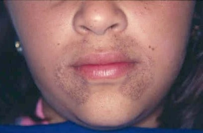
Lupus erythematosus affects the limbs, buttocks, areas under lupus lesions, and periarticular regions. Metastatic calcification produces periarticular lesions. Idiopathic calcification appears on children's faces as subepidermal calcified nodules, while iatrogenic calcification occurs at venipuncture sites.
Diagnosis
Dystrophic calcinosis cutis is the most common type; the calcified mass consists of amorphous calcium phosphate and hydroxyapatite. It is linked to connective tissue diseases, infections, hereditary disorders, and cutaneous neoplasms. Clinical presentation ranges from an incidental radiographic finding to subcutaneous nodules or plaques. Metastatic calcinosis cutis results from calcium salt precipitation in normal tissue due to abnormal phosphate or calcium metabolism; chronic renal failure is the most frequent cause.
Iatrogenic calcinosis cutis presents as firm dermal or subcutaneous nodules, often a warm, tender swelling at venipuncture sites after extravasation of intravenous calcium chloride, calcium gluconate, or phosphate-containing solutions; it can also appear as soft yellow-white epidermal plaques from electrode compounds. Traumatic calcinosis cutis occurs from occupational exposure, as in oil-field workers and coal miners. Idiopathic calcification is not linked to systemic illness or tissue injury and is usually localized, though one exceptionally broad case exists.
Idiopathic scrotal calcinosis affects young males, appearing as asymptomatic yellowish nodules 1 mm to several centimeters on the scrotum with normal blood calcium and phosphate levels.
Treatment
Diltiazem, a calcium channel blocker, is a mainstay of medical treatment; it is thought to modify intracellular calcium levels, reducing the capacity for calcium nidus production and crystallization. Colchicine, an antimicrotubule drug with anti-inflammatory properties, has been used for calcinosis cutis inflammation caused by a foreign body-like response, though with varying success.
More in Skin Conditions Resulting from Errors in Metabolism
Sources
Compiled from Wikipedia and the sources listed below. Text from Wikipedia is available under CC BY-SA 4.0; this entry is adapted from it.
- Wikipedia: Calcinosis cutis (CC BY-SA 4.0).
- Word definitions: the , each quoted from its Wikipedia article.
Spotted an error? Know more?
Reader corrections go straight into our review queue. Suggest an edit · How this site is sourced