Medical ultrasound
Uses high-frequency sound waves for medical imaging and therapy.
Last updated

Harrison Keely · CC BY 4.0
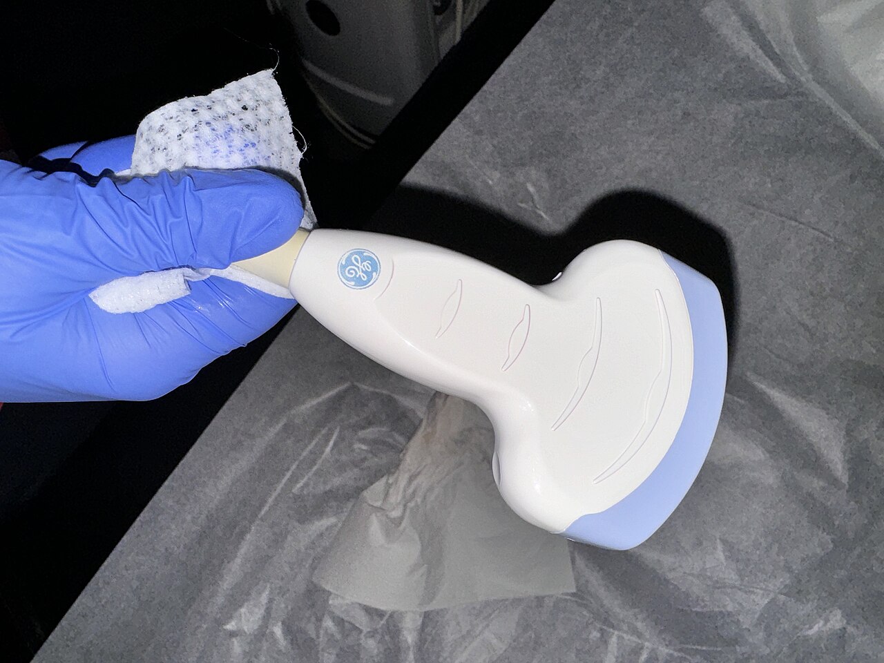
Medical ultrasound uses sound waves above 20,000 Hz to create images called sonograms by sending pulses into tissue with a probe.[1] Sonography is widely used for both diagnosis and therapeutic procedures, such as guiding biopsies or draining fluid collections.[2] Sonographers perform scans that are traditionally interpreted by radiologists or cardiologists.[2]
Quick Facts
- Field
- Medical imaging and therapy
- Types
- Diagnostic (sonography) and therapeutic
- Common modes
- A-mode
- B-mode
- M-mode
- 3D imaging
- Key techniques
- Doppler sonography
- harmonic imaging
- B-flow
Facts from the source article.
Key facts and history
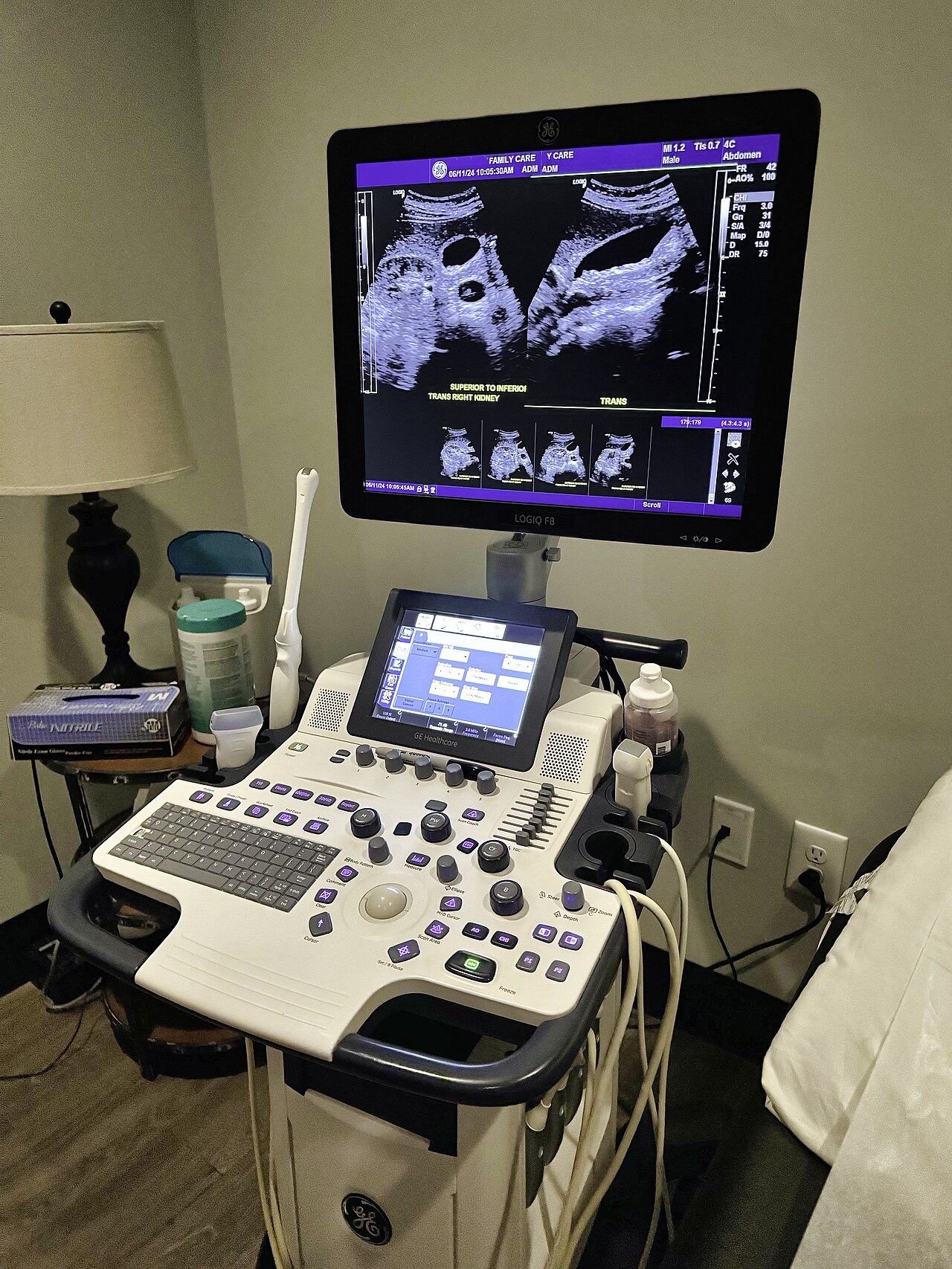
Ultrasound images are not true tomographic representations because the actual speed of sound varies greatly in different tissue types.[3] A-mode records transducer voltage amplitude as a function of two-way travel time of an ultrasound pulse.[4] To calculate imaging depth from two-way travel time, the majority of devices assume a speed of sound equal to 1540 m/s.[3] Volumetric imaging is achieved by merging B-mode scans obtained with either rotating or stationary specialized probes.[5]
Without requiring magnets or ionizing radiation, a combined rotating ultrasound and photoacoustic system generates three-dimensional views of both blood and tissue.[6][7] A volumetric picture is produced by this system through the use of a detector arc.[6] Hemoglobin molecules, set into vibration by a laser pulse, produce an acoustic signal that the detectors then absorb.[6][7] To position a treatment probe, A-mode and B-mode are commonly employed, often while therapy is being delivered.[8]
Ian Donald is credited with the initial development of obstetrical sonography during the late 1950s and 1960s.[9]
What the cited sources add
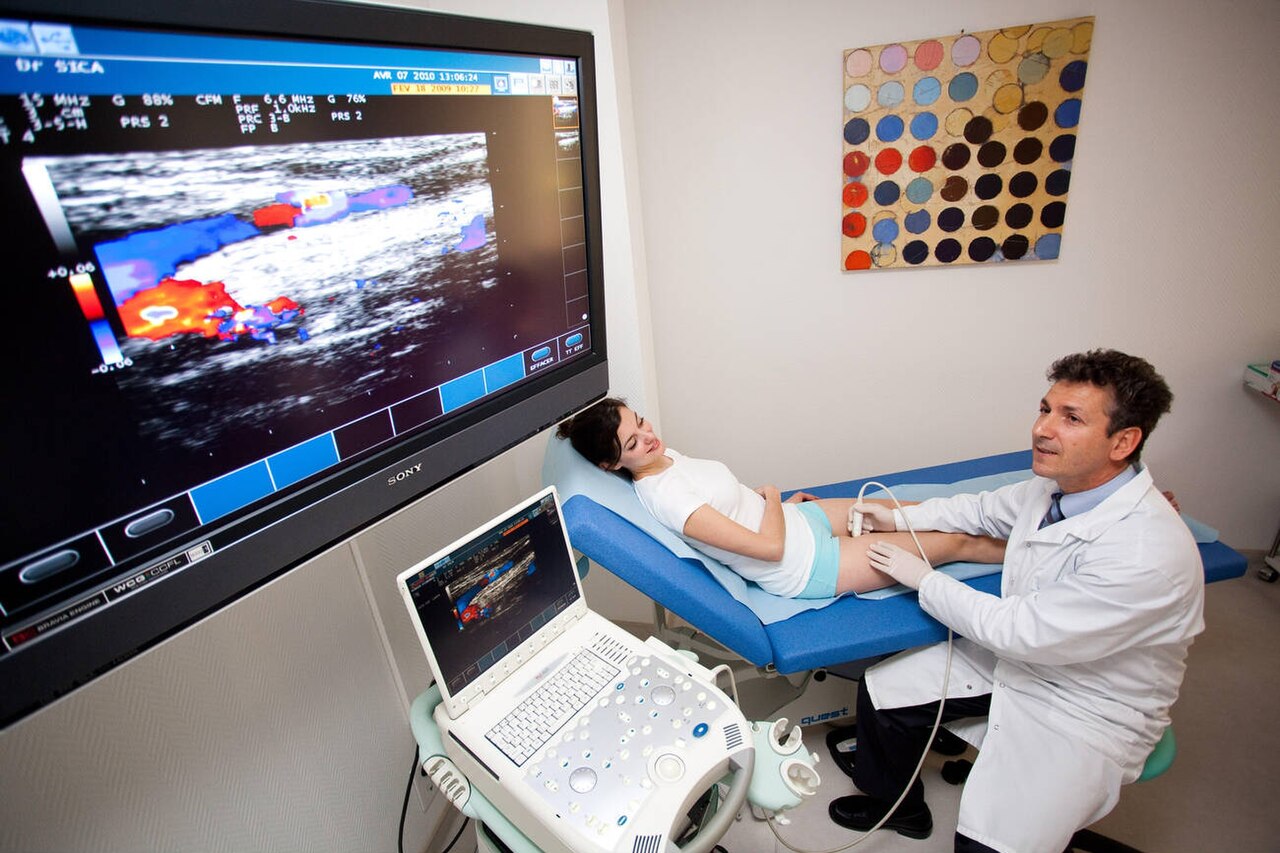
The British Medical Ultrasound Society states that a pelvic ultrasound scan is the most effective imaging modality to examine the uterus and ovaries.[10] The same source notes that transvaginal ultrasound is usually employed to examine the ovaries, pelvis, or part of the womb.[10] The USCOM 1A device is described as an advanced non-invasive Doppler ultrasound device for rapid optimization of the circulation.[11] The RUS-PAT system is less expensive to build than an MRI scanner and avoids radiation needed for X-ray and CT scans.[7]
A 2023 systematic review by Stone et al. found that ultrasound greatly assists catheter insertion into veins in challenging situations.[12][13]
References
- Wikipedia, "Medical ultrasound" (CC BY-SA 4.0), which cites: Aldrich, "Basic physics of ultrasound imaging", Critical Care Medicine, May 2007, p. S131–S137 (original not read by us) en.wikipedia.org
- Wikipedia, "Medical ultrasound" (CC BY-SA 4.0), which cites: Izadifar, "Mechanical and Biological Effects of Ultrasound: A Review of Present Knowledge", Ultrasound in Medicine & Biology, June 2017, p. 1085–1104 (we also read that source) en.wikipedia.org
- Wikipedia, "Medical ultrasound" (CC BY-SA 4.0), which cites: "EFSUMB Course Book", EFSUMB, 2019, p. 1–23 (original not read by us) en.wikipedia.org
- Wikipedia, "Medical ultrasound" (CC BY-SA 4.0), which cites: "Fundamentals of Medical Ultrasonics", CRC Press, 2011; The Gale Encyclopedia of Medicine, 2nd Edition, Vol. 1 A-B. p. 4; Cobbold, Richard S. C., "Foundations of Biomedical Ultrasound", Oxford University Press, 2007, p. 422–423 (we also read that source) en.wikipedia.org
- Wikipedia, "Medical ultrasound" (CC BY-SA 4.0), which cites: Cobbold, Richard S. C., "Foundations of Biomedical Ultrasound", Oxford University Press, 2007, p. 422–423 (we also read that source) en.wikipedia.org
- Wikipedia, "Medical ultrasound" (CC BY-SA 4.0), which cites: LeBlanc, "New 3D imaging system could address limitations of MRI, CT and ultrasound", Newsroom, 2026-01-16 (we also read that source) en.wikipedia.org
- LeBlanc, "New 3D imaging system could address limitations of MRI, CT and ultrasound", Newsroom, 2026-01-16. facts only, no text reused. keck.usc.edu
- Wikipedia, "Medical ultrasound" (CC BY-SA 4.0), which cites: Tzou, "Ultrasound Use in Urinary Stones: Adapting Old Technology for a Modern-Day Disease", Journal of Endourology, April 2017, p. S–89–S-94 (original not read by us) en.wikipedia.org
- Wikipedia, "Medical ultrasound" (CC BY-SA 4.0), which cites: Hellman, "Safety of Diagnostic Ultrasound in Obstetrics", The Lancet, May 1970, p. 1133–1135; Campbell, "A short history of sonography in obstetrics and gynaecology.", Facts, Views & Vision in ObGyn, 2013, p. 213–29 (original not read by us) en.wikipedia.org
- "Pelvic / Gynaecology Ultrasound (including transvaginal)", The British Medical Ultrasound Society. facts only, no text reused. bmus.org
- "Applications » Uscom". facts only, no text reused. uscom.com.au
- Wikipedia, "Medical ultrasound" (CC BY-SA 4.0), which cites: Stone, "Educational programs for implementing ultrasound guided peripheral intravenous catheter insertion in emergency departments: A systematic integrative literature review", Australasian Emergency Care, September 2023 (we also read that source) en.wikipedia.org
- Renee Stone, "Educational programs for implementing ultrasound guided peripheral intravenous catheter insertion in emergency departments: A systematic integrative literature review", Australasian Emergency Care (2023), doi:10.1016/j.auec.2023.06.001. OpenAlex metadata CC0; paper not read. doi.org
Each numbered sentence above was checked against the source it cites before publication. Sentences citing Wikipedia adapt text from the English Wikipedia article Medical ultrasound (contributors), used under CC BY-SA 4.0; this entry is available under the same licence. Other sources were used for facts only; no text or images are copied from them.
More in Science & Measurement
Sources
Compiled from Wikipedia and the sources listed below. Text from Wikipedia is available under CC BY-SA 4.0; this entry is adapted from it.
- Wikipedia: Medical ultrasound (CC BY-SA 4.0).
Spotted an error? Know more?
Reader corrections go straight into our review queue. Suggest an edit · How this site is sourced