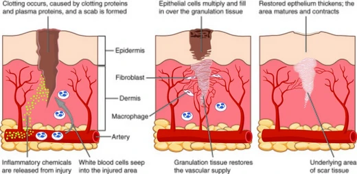
Healing of periapical lesions
Healing requires decontamination of the root canal system.

Healing of periapical lesions is a process that follows the removal of microbial infection from the root canal system. The body's inflammatory response, involving neutrophils and later macrophages and lymphocytes, can lead to tissue breakdown, but proper chemomechanical debridement and decontamination of the root canal system can allow resolution of the periapical lesion.
- Field
- Endodontics
- Known for
- Healing of periapical lesions through decontamination of the root canal system
Lore & Background
Healing of periapical lesions depends on obtaining and maintaining a decontaminated root canal system. The root canal system is not simply a tapered cone but can be an intricate labyrinth of anastomosing canals. Files produce shape, but irrigants clean the system; copious sodium hypochlorite is necessary to dissolve pulp tissue and destroy microorganisms. Conventional endodontic therapy, including chemical and mechanical debridement, is essential to disrupt the microbial ecosystem associated with the disease process. Recent studies have shown that intracanal application of chlorhexidine gluconate and calcium hydroxide prior to completing therapy can produce highly favorable results, even for large periapical lesions.
Reader's Guide
Healing of periapical lesions is achieved by removing the cause of infection, primarily through chemomechanical debridement of infected root canals. The presence of bacteria is the determinant for pulpal and periapical disease, as demonstrated by studies in germ-free rats. Endodontic infections are polymicrobial, and a positive correlation exists between the number of bacteria in an infected root canal and the size of the resultant periradicular radiolucency. Proper diagnosis and removal of the cause of infection, whether through endodontic therapy or extraction, allows for resolution of the periapical lesion. The traditional thought that endodontic therapy must be completed immediately is challenged by evidence that intracanal medicaments can lead to substantial healing before completion of therapy.
Did You Know?
- Intracanal application of chlorhexidine gluconate and calcium hydroxide was shown to lead to substantial healing of a large periapical lesion.
- The root canal system can be an intricate labyrinth of anastomosing canals, not merely a tapered cone.
Definition and Anatomical Context
The periapical cyst, widely recognized in dental literature as the most prevalent odontogenic cyst, occupies a well-defined anatomical niche. The term 'periapical' refers specifically to the tissues encircling the tip of a tooth's root, while 'cyst' designates a pathological cavity whose walls are lined by epithelium and whose interior holds fluid or gaseous material rather than accumulated pus. These lesions are exceptionally common in clinical practice, with a strong predilection for the maxillary anterior region. Their origin typically traces back to pulpal necrosis triggered by untreated dental caries or traumatic injury. The epithelial lining that characterizes the cyst derives from the rests of Malassez, small clusters of epithelial cells that normally reside near the root apex. Although a periapical cyst often begins without any noticeable symptoms, it carries considerable clinical weight because a secondary infection can introduce significant pain and progressive tissue damage. On standard radiographic imaging, the lesion presents as a radiolucent dark area encircling the root apex, providing dentists with a critical diagnostic landmark.
Three-Stage Developmental Pathway
Research describes the formation of a periapical cyst as unfolding across three distinct inflammatory stages. In the initial phase, the epithelial cells belonging to the rests of Malassez at the apex of a non-vital tooth—where the nerve and blood supply have already degenerated—receive stimulation from the body's inflammatory reaction to bacterial endotoxins or from direct contact with necrotic pulp tissue. This stimulus pushes the dormant cells back into an active growth phase, while bacterial byproducts simultaneously seep into the periapical region through the compromised pulp. During the second, development stage, those epithelial cells organize into strands that migrate toward areas of exposed connective tissue and foreign material. Multiple strands from individual rests converge and envelop the abscess or foreign body. The final growth stage is driven by fluid dynamics: because the osmolality inside the forming cavity exceeds that of the surrounding serum in nearby capillaries, fluid is drawn inward, progressively increasing both the internal pressure and the overall size of the cyst.
Clinical Progression and Complications
Periapical cysts typically begin as silent, slowly progressing lesions that a patient may not notice at all. The clinical picture changes dramatically once secondary infection takes hold, producing swelling and pain. Early in this symptomatic phase, the cyst manifests as a round, firm protrusion beneath the mucous membrane. Over time, however, the body resorbs portions of the cyst wall, transforming the structure into a softer, fluid-filled accumulation. As the lesion enlarges, it can expand surrounding bone, displace adjacent tooth roots, and even cause visible discoloration of the affected tooth. Diagnostic testing reveals a characteristic pattern: the tooth responds negatively to electric and ice vitality tests yet remains sensitive to percussion. Surrounding gingival tissue may show lymphadenopathy, and palpation of the alveolar plate can produce a crepitus sensation. The most serious complication arises when the cyst erodes through the floor of the maxillary sinus; once it enters the antrum, the available space accelerates its expansion rate, and tapping the affected teeth elicits sharp, shooting pain that is often clinically diagnostic of pulpal infection.
Competing Theories of Cyst Expansion
The precise mechanism driving periapical cyst growth remains a subject of active debate, with several competing theories offering partial explanations. The biomechanical theory attributes size increase to pressure and concentration gradients between the cystic cavity and its surroundings, which drive fluid movement inward. Two biochemical models add further nuance: one emphasizes collagenase-mediated breakdown of jaw bone collagen, releasing cytokines and growth factors that mobilize and proliferate epithelial cells; the other points to acidic metabolic products of the cyst itself—specifically Prostaglandin-2 and Interleukin-1—as agents that resorb bone and create additional space. A nutritional deficiency theory proposes that the innermost epithelial cells, being too distant from blood vessels, undergo ischemic liquefactive necrosis, and the resulting dead-tissue aggregate forms the cavity. Finally, the abscess theory holds that epithelial cells inherently reproduce to cover any exposed connective tissue, but an abscess must form first to trigger this response. Broadly, the field is divided between a complementary inflammatory response model and a chemical-reaction model centered on Interleukin and Prostaglandin activity.
Frequently Asked Questions
What is Healing of periapical lesions?
It is the biological resolution process in endodontics that occurs once microbial infection has been successfully removed from the root canal system. Rather than being a single event, it represents the body's gradual repair of the inflamed tissue surrounding the tooth's apex.
What drives the inflammatory response that creates the periapical lesion?
Neutrophils arrive first at the site of infection, followed by macrophages and lymphocytes, and this cascading immune activity can progressively break down the surrounding periapical tissue. The longer the microbial load persists, the deeper the tissue damage becomes.
What is the single most critical requirement for the lesion to actually heal?
Decontamination of the root canal system is non-negotiable; without thorough chemomechanical debridement that eliminates the microbial source, the inflammatory cycle simply continues. In other words, you cannot expect periapical resolution while the canal remains contaminated.
Which dental specialty deals with Healing of periapical lesions?
It sits squarely within endodontics, the branch of dentistry dedicated to the pulp, root canal system, and the tissues immediately surrounding the root apex. Endodontists are the ones who perform the debridement and decontamination that sets healing in motion.
How does the healing process resolve once treatment is complete?
After the root canal has been properly debrided and decontaminated, the stimulus for inflammation is removed, allowing neutrophils, macrophages, and lymphocytes to withdraw from the periapical area. The damaged tissues then undergo natural repair and the lesion gradually resolves over time.
More in Healing 1-19
Spotted an error? Know more?
Reader corrections go straight into our review queue. Suggest an edit · How this site is sourced