Craniofacial regeneration
Biological process of skull and face regrowth after injury.

Craniofacial regeneration is the natural process that allows the bones of the skull and face to regrow after an injury. This healing is needed when facial tissue is damaged, either from surgical procedures or trauma. Surgeons may intentionally fracture a patient’s face to correct conditions like cleft lip, Apert syndrome, Treacher Collins syndrome, Oligodontia, Cherubism, Crouzon syndrome, Pfeiffer syndrome, craniosynostosis, or Goldenhar syndrome. Other surgical applications include fixing birth defects such as hypertelorism, performing maxillofacial surgery, treating rare craniofacial clefts, or removing tumors. Outside of surgery, facial injuries often result from automotive accidents. Most craniofacial defects are present at birth, occurring in about 1 in 700 live births, which amounts to approximately 1.3 million children each year. Common corrective surgeries include intracranial procedures to expand the skull for brain growth, cleft palate repairs to close gaps in the roof of the mouth, and cleft lip surgeries to close gaps in the lip. Most people with these abnormalities have a normal life expectancy, though symptoms like abnormal skull shape, difficulty with breathing, hearing, swallowing, or speech, and facial paralysis can persist throughout life.
Research into craniofacial regeneration gained momentum after mesenchymal stem cells (MSCs) were discovered by A.J. Friedenstein in the 1970s. MSCs are found in many postnatal tissues, including orofacial tissues, and have shown the most promise for regeneration. Alginate hydrogel containing nerve growth factor has been used to deliver these stem cells to healing tissues. Although there are few craniofacial-specific clinical trials for stem cell therapies, scientists have identified several craniofacial-specific stem cell populations that can develop into various skull structures. These include bone marrow mesenchymal stem cells (BMMSCs), adipose-derived mesenchymal stem cells (AMCs), muscle satellite cells (MuSCs), periodontal ligament stem cells (PDLSCs), and stem cells from human exfoliated deciduous teeth (SHED). Two of the most promising types are BMMSCs and AMCs.
BMMSCs have been used to repair craniofacial defects.
- Field
- Regenerative medicine, craniofacial biology
- Known for
- Biological process of skull and face regrowth after injury
- Prevalence
- 1 in 700 live births (approximately 1.3 million children per year)
- Key cell types
- Bone marrow mesenchymal stem cells (BMMSC), adipose-derived mesenchymal stem cells (AMCs)
- Major funding
- $24 million awarded by NIDCR in 2017 to two research centers
Lore & Background
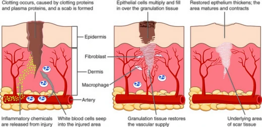
Craniofacial regeneration is initiated by an inflammatory response to injury, followed by angiogenesis, leading to mesenchymal stem cell (MSC) differentiation. During inflammation, macrophages recruit lymphocytes that secrete cytokines, which stimulate angiogenesis and MSC differentiation into osteoblasts. Angiogenesis, the formation of blood vessels from existing ones, allows oxygen, nutrients, and progenitor cells to reach the regeneration site. Vascular endothelial growth factor (VEGF) plays a crucial role in both angiogenesis and osteogenesis, and loss-of-function experiments against VEGF in osteoblast precursors significantly reduce ossification in craniofacial bone structures.
Reader's Guide
Craniofacial regeneration research has yielded promising results using mesenchymal stem cells (MSCs), discovered in the 1970s by A.J. Friedenstein. Bone marrow mesenchymal stem cells (BMMSCs) have been shown to repair craniofacial defects in animal models including canines, mice, and sheep. Adipose-derived mesenchymal stem cells (AMCs) showed success in a small clinical trial (n=13) where 10 out of 13 patients successfully integrated AMCs with scaffolds of bioactive glass or β-tricalcium phosphate. Despite a lack of craniofacial-specific clinical trials for stem cell therapies, significant effort has identified craniofacial-specific stem cell populations. The National Institute of Dental and Craniofacial Research awarded $24 million in 2017 to two centers focused on craniofacial disease and injury research. Most patients with craniofacial abnormalities have a normal life expectancy, though symptoms such as abnormal cranial morphology, difficulty in breathing, hearing, swallowing, or speech, or facial paralysis often persist throughout life.
Did You Know?
- Craniofacial regeneration is not to be confused with tooth regeneration.
- Chronic inflammation, which mimics aging, has been shown to negatively affect bone regeneration.
- In 1994, BMMSCs grown with dexamethasone, ascorbic acid 2-phosphate, and inorganic phosphate differentiated into functional osteoblast-like cells.
Clinical Scope & the Burden of Congenital Defects
Craniofacial regeneration serves as the biological foundation for a wide range of medical interventions. When surgeons deliberately fracture facial structures to address congenital conditions—such as cleft lip, Apert syndrome, Treacher Collins syndrome, Craniosynostosis, or Goldenhar Syndrome—the body must rebuild the affected bone and soft tissue. Beyond corrective surgery, this regenerative capacity is also called upon after maxillofacial tumor removal, repair of rare craniofacial clefts, correction of hypertelorism, and treatment of trauma most commonly caused by automotive accidents. The scale of need is substantial: craniofacial defects are congenital in the majority of cases, affecting roughly one in every 700 live births, which translates to an estimated 270,000 children annually worldwide. Standard corrective procedures span intracranial surgeries that create additional space for a developing brain, cleft palate operations that close gaps in the oral roof, and cleft lip repairs. Despite the severity of these conditions, most affected individuals maintain a normal life expectancy, though they often contend with lifelong challenges including abnormal skull shape, impaired breathing, hearing, swallowing, speech difficulties, or facial paralysis.
Stem Cell Therapies & the Road to Clinical Translation
The discovery of mesenchymal stem cells by A. J. Friedenstein in the 1970s opened a new frontier for craniofacial repair. Because MSCs reside in numerous postnatal tissues, including orofacial regions, they have become the most promising cellular tool for regrowing skull and facial bone. Researchers have catalogued several craniofacial-specific populations—bone marrow MSCs, adipose-derived MSCs, muscle satellite cells, periodontal ligament stem cells, and stem cells from human exfoliated deciduous teeth—each capable of giving rise to distinct skull structures. In 1994, BMMSCs cultured with dexamethasone, ascorbic acid 2-phosphate, and inorganic phosphate successfully differentiated into osteoblast-like cells, though in vivo testing showed only a little over half of treated mice developed bone structure. Adipose-derived MSCs fared better in the clinic: in 2014, George K. Sándor transplanted AMCs alongside bioactive glass or β-tricalcium phosphate scaffolds into 13 patients with craniomaxillofacial defects, achieving successful integration in 10. By 2017, the NIDCR committed $24 million to two centers dedicated to craniofacial disease and injury research, signaling growing institutional investment in this field.
The Regenerative Cascade: Inflammation, Angiogenesis, and Vascular Control
Craniofacial regeneration follows a tightly sequenced biological cascade. The process begins with an inflammatory response to tissue injury, during which collagen fibers in connective tissue are disrupted to release healing proteins. Macrophages are activated and recruit lymphocytes to the wound site; these lymphocytes secrete cytokines—proteins that mediate the immune response and, crucially, stimulate both angiogenesis and the differentiation of MSCs into osteoblasts, the cells that ultimately form new bone. A critical nuance emerges here: while acute inflammation is necessary, chronic inflammation, which mimics the effects of aging, has been shown to impair bone regeneration, though the precise threshold remains incompletely understood within the broader immune context. Following inflammation, angiogenesis takes over, generating new blood vessels through rapid outgrowth and organization. This vascular remodeling depends heavily on extracellular signals including cytokines, proteases, and growth factors. Transmembrane receptor proteins called integrins play a pivotal role; inhibiting integrin α5β1 halts angiogenesis entirely, and targeting integrin αvβ5 negatively affects VEGF-dependent vessel formation, underscoring how tightly regulated this vascular phase is.
Research Landscape, Delivery Systems, and Translational Gaps
The scientific study of craniofacial regeneration sits at the intersection of developmental biology, immunology, and regenerative medicine, yet it remains explicitly distinct from the separate field of tooth regeneration. Since Friedenstein's identification of mesenchymal stem cells in the 1970s, the field has expanded to encompass a diverse array of craniofacial-specific precursor populations, each with the potential to generate particular skull structures. Despite this breadth, a notable gap persists: there is a lack of craniofacial-specific clinical trials for stem cell therapies, meaning much of the evidence still rests on animal models in canines, mice, and sheep. Alginate hydrogel containing nerve growth factor has been explored as a delivery vehicle for stem cells during the regenerative process. The 2014 Sándor trial, with its modest cohort of 13 patients and 10 successful integrations, represents one of the few human data points in the literature. The 2017 NIDCR award of $24 million to two dedicated research centers reflects both the promise and the unfinished work in this domain, as scientists continue to bridge the distance between promising in-vitro differentiation and reliable in-vivo bone reconstruction.
Frequently Asked Questions
What is Craniofacial regeneration?
Craniofacial regeneration is the body's innate biological ability to regrow skull and facial bones after they have been damaged by trauma or surgery. It sits at the intersection of regenerative medicine and craniofacial biology.
What conditions does Craniofacial regeneration help correct?
Surgeons intentionally fracture facial bones so this regrowth process can reshape them when treating cleft lip, craniosynostosis, Apert syndrome, Treacher Collins syndrome, and other congenital or traumatic deformities. It also underpins maxillofacial surgery and repair of rare craniofacial clefts.
How prevalent is the need for Craniofacial regeneration?
Roughly one in every 700 live births involves a craniofacial condition that depends on this regenerative response, amounting to approximately 1.3 million children per year worldwide.
What are Craniofacial regeneration's key cell types?
The process is driven primarily by bone marrow mesenchymal stem cells (BMMSC) and adipose-derived mesenchymal stem cells (AMCs), which supply the new bone-forming cells needed to rebuild the skull and face.
What major funding has supported Craniofacial regeneration research?
In 2017, the NIDCR granted $24 million to two dedicated research centers to deepen understanding of how skull and facial bones regenerate after injury.
More in Healing 1-19
Spotted an error? Know more?
Reader corrections go straight into our review queue. Suggest an edit · How this site is sourced