
Constriction ring syndrome
Congenital disorder with unknown cause, producing constriction rings on extremities.

Fyrosth · CC0
Constriction ring syndrome (CRS) is a birth defect of unknown cause. It happens when bands or rings form inside the womb, creating deep grooves—most often in the fingers and toes. In rare cases, these rings can wrap around other parts of the fetus, leading to amputation or even death before birth. The parts of the body above the constriction or amputation develop normally.
CRS can occur alongside other malformations, with club foot being the most common. The exact shape of the bands, the amount of swelling, and the type of amputation vary from person to person and cannot be predicted. Usually, more than one limb is affected, and it is unusual for a single ring to appear alone without any other signs of the syndrome.
**Signs and symptoms** Constriction by amniotic bands can cause: - Rings around fingers, arms, or legs - Swelling beyond the constriction point (congenital lymphedema) - Amputation of fingers, arms, or legs (congenital amputation)
**Cause** There are three main theories about what causes CRS.
The first theory, proposed by Streeter in 1930, suggests an intrinsic problem with the germ plasm. This idea is supported by the fact that constriction rings often appear with other internal and systemic anomalies. Because of these additional defects, the condition is sometimes called constriction ring syndrome, constriction band syndrome, or Streeter bands. Some researchers link this to a shared vascular disruption that also occurs with cleft palate and other cleft defects, pointing to a possible circulation problem.
The second theory involves an intrauterine disruption, such as the amnion rupturing early in the second trimester. When the amnion separates from the chorion, it creates thin strands that can entangle the fingers or toes. As the fetus grows but the strands do not, they tighten, reducing blood flow and causing abnormalities. In some cases, a limb or digit may be completely amputated before birth, or it may die and require surgical removal after birth. Names like amniotic band syndrome (ABS), amniotic disruption complex, and amniochorionic mesoblastic fibrous strings come from this theory.
The third theory points to intrauterine trauma, such as from amniocentesis or fetal surgery. This trauma could cause bleeding that leads to acrosyndactyly. One study confirmed the presence of bands on ultrasound after fetal surgery.
- Field
- Congenital disorder
- Known for
- Constriction rings around digits, arms, and legs; congenital lymphedema; congenital amputation
- Associated malformations
- Club foot most common; also clubhands, cleft lip, cleft palate, hemangioma
- Classification system
- Patterson classification (four categories: simple rings, rings with deformity, acrosyndactyly, uterine amputation)
Lore & Background
The cause of constriction ring syndrome is disputed, with three main theories. The intrinsic theory, proposed by Streeter in 1930, implicates an anomaly in germ plasm, reinforced by the presence of other internal visceral anomalies. The second theory involves intrauterine disruption from spontaneous amniotic rupture early in the second trimester, producing strands that entangle digits and constrict as the fetus grows. The third theory postulates intrauterine trauma, such as amniocentesis or fetal surgery, leading to hemorrhage and acrosyndactyly. Because of these differing theories, the syndrome has many names, including amniotic band syndrome (ABS), Streeter bands, and amniochorionic mesoblastic fibrous strings.
Reader's Guide
Constriction ring syndrome is significant as a complex, asymmetric congenital anomaly with highly variable manifestations. Diagnosis is often confirmed via ultrasonography, though individual strands are small and hard to see, so bands are detected indirectly through constrictions and swelling. Misdiagnosis is common, and detailed ultrasound or MRI may be needed. The Patterson classification system, based on severity, guides treatment: simple rings, rings with distal deformity or lymphedema, rings with acrosyndactyly, and uterine amputation. The syndrome's legacy lies in its unresolved etiology—no single theory explains all cases—and in its clinical variability, where no two cases are exactly alike. The condition underscores the importance of careful prenatal imaging and the need for individualized surgical planning.
Did You Know?
- More than one extremity is usually affected, and it is rare for only one ring to present as an isolated malformation.
- 31.5% of clubfoot cases can be correlated with ABS, with 20% occurring bilaterally.
- The thumb is not likely to be affected because the fetus typically holds it in tight adduction flexion.
- Patterson classification includes four categories: simple constriction rings, rings with deformity, acrosyndactyly, and uterine amputation.
The Three Competing Theories of Origin
Despite decades of research, no single accepted explanation exists for why constriction ring syndrome develops in a fetus. Three competing theories have emerged, each generating its own cluster of names for the condition. The first, proposed by Streeter in 1930, attributes the defect to an inherent anomaly in germ plasm. This intrinsic view is bolstered by the frequent co-occurrence of the rings with internal visceral anomalies and by the observed link to vascular disruption seen in cleft palate cases. The second theory is mechanical: spontaneous rupture of the amnion early in the second trimester separates it from the chorion, leaving behind thin strands that can snare digits or toes. As the fetus grows while the bands stay fixed, they tighten, cutting off circulation and potentially producing a natural amputation or necrotic tissue requiring surgical removal after birth. The third theory points to intrauterine trauma—amniocentesis or fetal surgery—as a trigger, with one study confirming bands via sonography after such a procedure. Critically, research has shown that not every affected child had a genuine amniotic strand, meaning the mechanical explanation alone cannot account for all cases. This unresolved etiology is why the condition has accumulated so many different and sometimes inaccurate names.
A Syndrome That Defies Prediction
Constriction ring syndrome is, by its very nature, a condition that resists generalization. The precise configuration of the bands, the degree of lymphedema, and the character of any resulting amputation vary from one patient to the next, making it impossible to predict what a given case will look like. The malformation most commonly produces deep grooves in the distal extremities—fingers and toes—but in rarer instances the constriction can encircle other parts of the fetus, leading to amputation of entire limbs or even intrauterine death. What remains consistent is that the anatomy proximal to the site of constriction or amputation is developmentally normal. Swelling of the extremities distal to the ring, known as congenital lymphedema, is a frequent companion, and amputation of digits, arms, or legs represents the most severe endpoint. More than one extremity is usually affected, and it is rare for a single ring to present as an isolated malformation with no other manifestation of the syndrome. The condition is also frequently accompanied by other structural anomalies, with club foot being the most common association. In short, no two cases of constriction ring syndrome are exactly alike, a fact that complicates both diagnosis and treatment planning.
The Diagnostic Maze
Identifying constriction ring syndrome before birth is possible but far from straightforward. Ultrasonography can confirm the diagnosis, sometimes as early as the end of the first trimester or the beginning of the second, yet most patients are not diagnosed until birth. The fundamental difficulty is that individual amniotic strands are tiny and nearly invisible on standard ultrasound; bands are detected indirectly through the constrictions and swelling they produce on limbs or digits. Misdiagnosis is common, so clinicians are advised to pursue further detailed imaging whenever signs of amniotic bands appear. Three-dimensional ultrasound and magnetic resonance imaging offer more precise visualization of the bands and the extent of danger to the fetus. Decreased fetal movement can be a warning sign of a serious problem, including amniotic band involvement. In rare but devastating cases, the membrane can wrap around the placenta or the baby's neck, causing strangulation and death. The differential diagnosis is equally challenging, as conditions such as symbrachydactyly, congenital amputations, digital hypoplasia, Adams–Oliver syndrome, and ADAM complex can all mimic or be confused with constriction ring syndrome. Adams–Oliver syndrome, in particular, is often mislabeled as CRS, yet it involves cutis aplasia of the scalp and digital hypoplasia without the characteristic constriction rings or edema.
Classification, Naming, and the Broader Clinical Picture
Because no two cases of constriction ring syndrome are exactly alike, creating a workable classification has always been difficult. The most widely used system, proposed by Patterson, is organized around the severity of the syndrome—a practical distinction because different severity levels call for different treatments. Other clinicians have refined this scheme by subdividing the depth of the ring into mild, moderate, severe, and amputation, and by further noting the presence or absence of lymphedema or soft tissue loss distal to the ring. The naming landscape is just as tangled. Depending on which etiological theory is favored, the condition may be called constriction ring syndrome, constriction band syndrome, Streeter bands, amniotic band syndrome, amniotic disruption complex, or amniochorionic mesoblastic fibrous strings. The clinical picture extends well beyond the rings themselves. There is a strong relationship between the syndrome and clubfoot: 31.5 percent of clubfoot cases can be correlated with amniotic band involvement, and 20 percent of those occur bilaterally. Other associated abnormalities include clubhands, cleft lip, cleft palate, and hemangioma. This constellation of asymmetric congenital anomalies, combined with the unknown cause and the proliferation of names, makes constriction ring syndrome one of the more complex conditions in congenital medicine.
Gallery
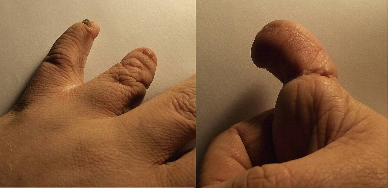
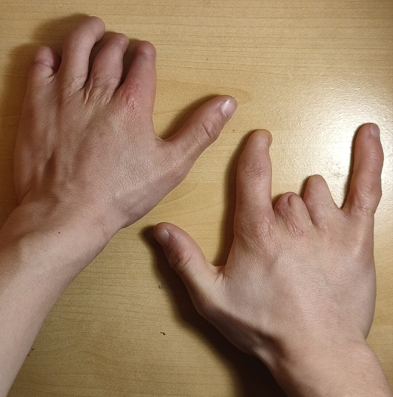
Frequently Asked Questions
What is Constriction Ring Syndrome and what makes it unique among congenital disorders?
CRS is a birth defect in which fibrous bands form around a developing fetus in utero, carving deep groove-like rings—most often on fingers and toes. Unlike many other genetic conditions, no specific gene has ever been linked to it, and its cause remains officially unknown.
What does Constriction Ring Syndrome actually look like on a patient?
The hallmark finding is one or more circumferential constrictions that create a visible ring or groove around a digit, limb, or occasionally the torso. Tissue above the ring typically develops normally, while the area below may show swelling, deformity, or complete amputation depending on severity.
How do clinicians categorize the different presentations of Constriction Ring Syndrome?
The Patterson classification sorts cases into four tiers: simple rings, rings accompanied by deformity, acrosyndactyly (fused digits), and uterine amputation. This framework helps doctors gauge severity and plan surgical or rehabilitative intervention.
What other malformations can appear alongside Constriction Ring Syndrome?
Club foot is by far the most common co-occurring anomaly, but cleft lip, cleft palate, hemangioma, and clubhands have all been documented in affected individuals. The exact constellation varies widely from one patient to the next and cannot be reliably predicted.
Why does Constriction Ring Syndrome remain a mystery in genetics despite having an OMIM entry?
Although it is catalogued in the Online Mendelian Inheritance in Man database, no causative gene or chromosomal locus has been identified to date. The condition is thought to arise from mechanical or vascular disruption during fetal development rather than from a single inherited mutation, which is why it resists the usual gene-mapping approach.
More in Genetic disorders with OMIM but no gene 1-24
Spotted an error? Know more?
Reader corrections go straight into our review queue. Suggest an edit · How this site is sourced