
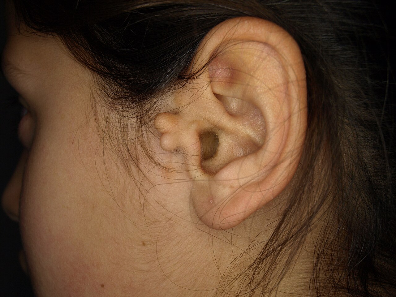
Accessory auricle
Congenital nodule near the ear, often surgically correctable.

Klaus D. Peter, Wiehl, Germany · CC BY 3.0 de
An accessory auricle is a congenital condition that appears as a small, skin-colored bump, most often located just in front of the ear. It can occur on one or both sides and may also appear elsewhere around the ear. The bump may be flat or stalk-like, soft or firm, and can contain cartilage. Under a microscope, the tissue resembles a normal outer ear, with skin and cartilage, though cartilage is not always present. Some researchers believe the tragus comes from the first branchial arch, suggesting that true accessory auricles are duplications of hillocks from the second branchial arch, appearing as a mirror-image ear folded forward on the cheek.
This condition is different from squamous papilloma and benign teratoma. It ranges from a simple ear tag or preauricular tag to a supernumerary ear (polyotia). It is a common anomaly of the first or second branchial arches and may occur alongside cleft palate, cleft lip, or underdeveloped lower jaw. It is also linked to Goldenhar syndrome and Wildervanck syndrome, and possibly to a congenital cartilaginous rest of the neck.
Simple surgical removal cures the condition. The recommended approach is to peel away the skin from the extra tissue and trim any protruding cartilage, which usually restores a normal appearance. Reconstruction is successful for true accessory auricles and for auricular appendages.
These lesions are usually noticed in newborns, though some people seek treatment later in life for cosmetic reasons. They affect males and females equally and occur in about 3 to 6 per 1,000 live births. A study of one family with 11 affected members found the condition was inherited in an autosomal dominant pattern.
Quick Facts
- Field
- dermatology
Facts from the source article.
Lore & Background
The general presentation of an accessory auricle is a skin-covered nodule, papule, or nodule, usually immediately anterior to the auricle, though it may appear anywhere within the periauricular tissues. Bilateral presentation can occur. Histologic examination shows it is a recapitulation of the normal external auricle, with skin, cartilaginous structures, and sometimes cartilage.
Reader's Guide
Accessory auricle is a congenital anomaly of the first or second branchial arches, presenting as a nodule near the ear. It is typically diagnosed at birth but may not be noticed until adulthood for cosmetic reasons. Simple surgical excision is curative, with normal appearance achieved in most cases. The condition may be associated with other anomalies such as cleft palate, cleft lip, or mandibular hypoplasia, and with syndromes like Goldenhar syndrome and Wildervanck syndrome. Its significance lies in its common occurrence and straightforward management, though it can indicate broader developmental issues.
Did You Know?
- Accessory auricle is present in approximately 3–6 per 1000 live births.
- A study of a family with 11 affected individuals showed autosomal dominant inheritance.
- The lesion may be soft or have a cartilaginous structure.
- It is associated with Goldenhar syndrome and Wildervanck syndrome.
Clinical Presentation & the Spectrum of Development
An accessory auricle most commonly manifests as a small, skin-coloured nodule situated just in front of the ear, though it can appear anywhere within the periauricular region. The lesion may be sessile or pedunculated, and its texture ranges from soft tissue to a firmer cartilaginous structure. In some individuals, the anomaly presents bilaterally, affecting both sides of the face. Clinically, the spectrum of development is broad: at the mildest end, it appears as a simple ear tag or preauricular appendage, while at the most extreme, it takes the form of a fully formed supernumerary ear, a condition termed polyotia. Histologically, the tissue recapitulates the architecture of a normal external ear, containing skin, cartilaginous elements, and in many variants, true cartilage. Importantly, these structures are distinctly different from squamous papillomas or benign teratomas, a distinction that guides accurate diagnosis and appropriate management.
Embryological Origins & Hereditary Pattern
Understanding the developmental biology behind an accessory auricle reveals its true nature. The tragus is considered the only hillock derived from the first branchial arch, which means that genuine cases of accessory auricle represent a duplication of the hillocks belonging to the second branchial arch. In its most complete form, the extra ear appears as a mirror image folded forward and resting on the posterior cheek. Histologic examination confirms this, showing the same layered components—skin, cartilaginous structures, and cartilage—found in a normal external auricle, though cartilage is absent in some variants. On the genetic side, a family study involving eleven affected members demonstrated that the trait follows an autosomal dominant pattern of inheritance, suggesting a single gene variant can be passed from parent to child. This congenital anomaly of the first or second branchial arches thus has a clear hereditary component in at least some families.
Associated Conditions & Diagnostic Context
Although an accessory auricle is often an isolated finding, its presence can signal a broader constellation of developmental anomalies. Concurrent conditions that may accompany it include cleft palate, cleft lip, and mandibular hypoplasia, all pointing to disrupted craniofacial development during the embryonic period. Two named syndromes carry a recognized association: Goldenhar syndrome, also known as oculo-auriculo-vertebral syndrome, and Wildervanck syndrome. Additionally, a link exists with congenital cartilaginous rests of the neck, further underscoring the connection to branchial arch development. From a diagnostic standpoint, the accessory auricle must be carefully distinguished from squamous papilloma and benign teratoma, as these conditions can superficially resemble the nodule or papule of an accessory structure. The histologic examination—revealing the characteristic triad of skin, cartilaginous tissue, and cartilage—serves as the definitive tool for separating a true accessory auricle from its mimics and for confirming the degree of structural development present.
Surgical Correction & Population Prevalence
From a treatment perspective, an accessory auricle is remarkably straightforward to correct. Simple surgical excision is curative: the overlying skin is peeled away from the extra-auricular tissue, and any protruding cartilage remnants are trimmed. In the majority of cases, this procedure restores a normal cosmetic appearance. Reconstruction outcomes are successful not only in true accessory auricle cases but also in individuals presenting with simpler auricular appendages. Epidemiologically, these lesions are present at birth, though they may not attract clinical attention until adulthood, when cosmetic concerns prompt a visit to a physician. There is no gender predilection—males and females are equally represented. The incidence is estimated at roughly three to six cases per one thousand live births, making it a relatively common congenital anomaly. Despite its frequency, the condition is benign and, once addressed surgically, carries no long-term sequelae.
Gallery
Frequently Asked Questions
What is accessory auricle and where does it show up on the body?
It is a congenital, skin-colored bump that typically sits just in front of the ear, though it can appear on one or both sides or at other spots around the ear. The nodule may be flat or on a small stalk, feel soft or firm, and sometimes contain a bit of cartilage.
Does accessory auricle have a known gene or a clear inheritance pattern?
No single gene has been assigned to it in OMIM, which is why it sits in the 'no gene' section of the encyclopedia. In one well-studied family the trait followed an autosomal-dominant pattern, but broader genetic underpinnings remain unresolved.
How common is accessory auricle and does it favor one sex?
Roughly 3 to 6 out of every 1,000 live births are affected, making it one of the more frequently noted minor ear anomalies. There is no reported gender predilection, so it shows up at similar rates in males and females.
What does the bump actually look like under a microscope, and where does the tissue come from developmentally?
Histologically the nodule mirrors a normal outer ear, displaying epidermis and, when present, cartilage. Developmentally, researchers link true accessory auricles to duplicated hillocks of the second branchial arch, distinguishing them from the tragus, which derives from the first arch.
Is accessory auricle ever a sign of something bigger, and how is it dealt with?
When it appears alongside other features it can be a marker for Goldenhar syndrome or Wildervanck syndrome, so clinicians look for associated findings. In isolated cases the standard treatment is a straightforward surgical excision, which is usually curative.
More in Genetic disorders with OMIM but no gene 1-24
Spotted an error? Know more?
Reader corrections go straight into our review queue. Suggest an edit · How this site is sourced