Branchial cleft cyst
Congenital neck cyst from failed embryonic cleft obliteration.

Ed Uthman from Houston, TX, USA · CC BY 2.0
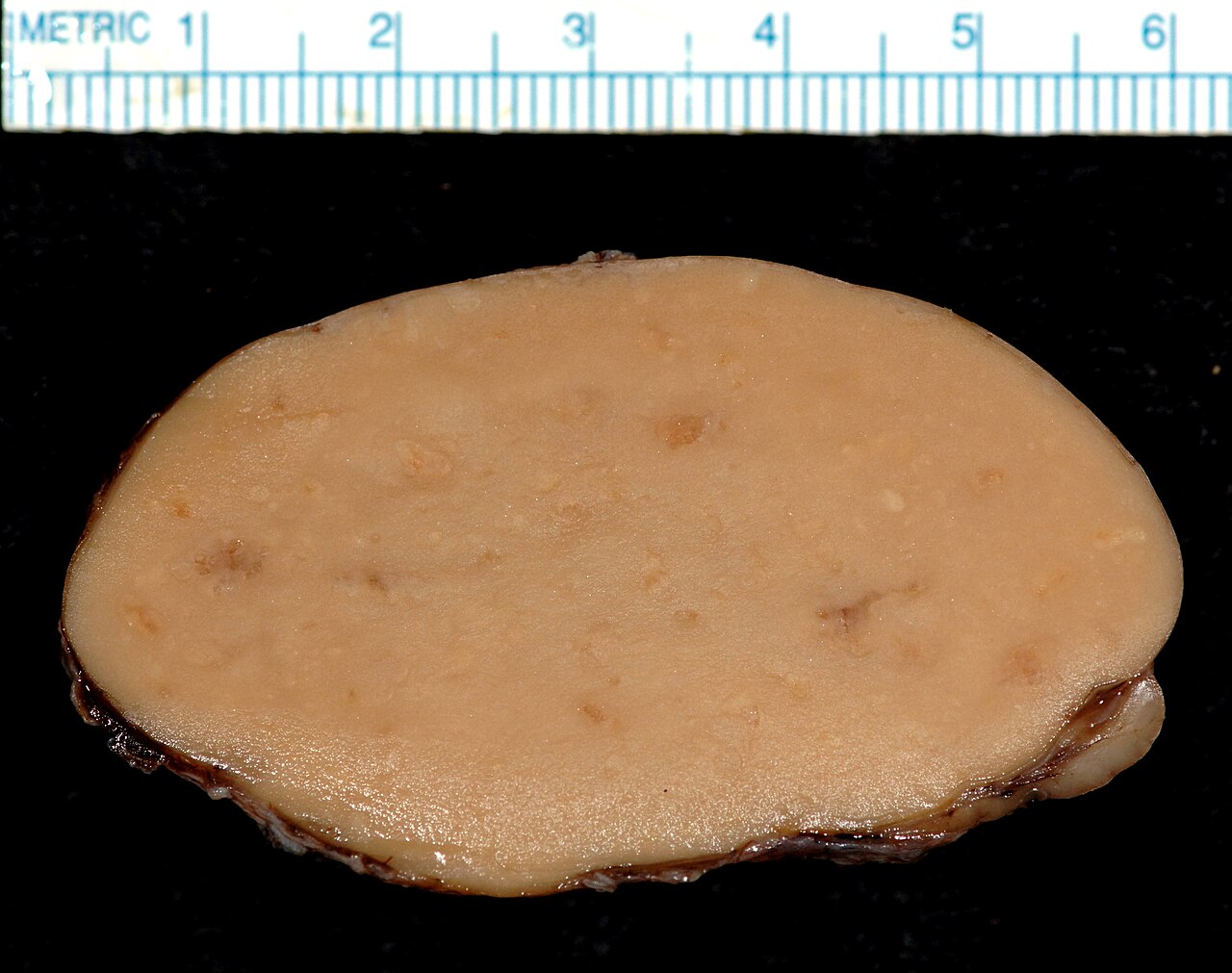
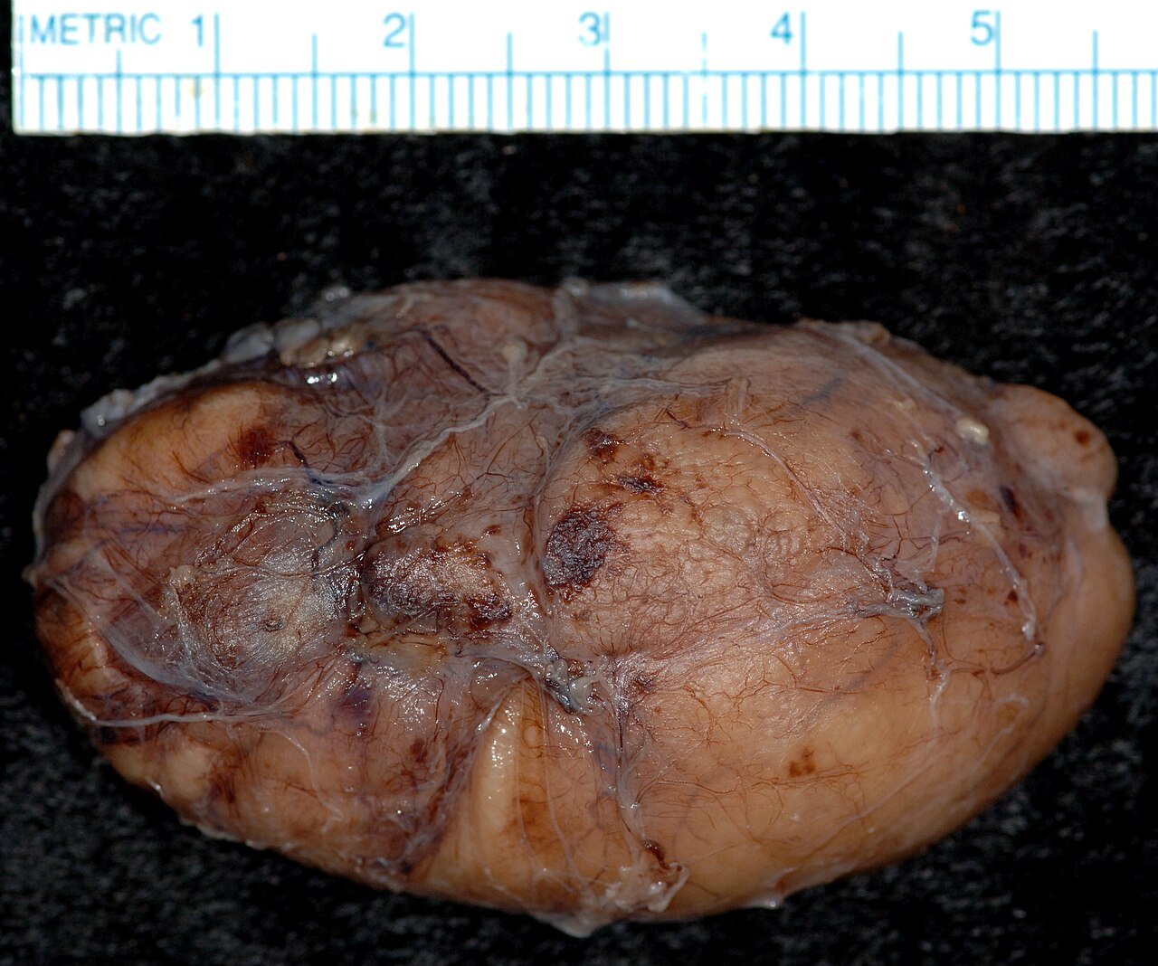
A branchial cleft cyst is a swelling in the upper part of the neck, typically located anterior to the sternocleidomastoid muscle. It may or may not have an opening to the skin surface, called a fistula. The condition arises from a developmental abnormality in the early prenatal period, usually due to failure of obliteration of the second, third, or fourth branchial cleft, or failure of fusion of the second branchial arches and epicardial ridge. Branchial cleft cysts account for almost 20% of neck masses in children.
Quick Facts
- Symptoms
- Painless, firm mass lateral to midline, usually anterior to the SCM, which does not move with swallowing
- Causes
- Family history
- Treatment
- Conservative, surgical excision
Facts from the source article.
Lore & Background
Branchial cleft cysts are remnants of embryonic development, resulting from a failure of obliteration of one of the four branchial clefts that form during human embryogenesis. These clefts are homologous to structures that develop into gills in fish. The most common type, second branchial cleft cysts, account for 90 to 95% of neck cysts and are located anterior to the sternocleidomastoid muscle, posterior to the submandibular gland, and lateral to the carotid sheath. First branchial cleft cysts, also called periauricular cysts, are always in or adjacent to the parotid gland and account for 8% of neck sinuses and cysts. Third and fourth branchial cleft anomalies are rare, with fourth cleft abnormalities presenting as sinus tracts or fistulas rather than true cysts.
Reader's Guide
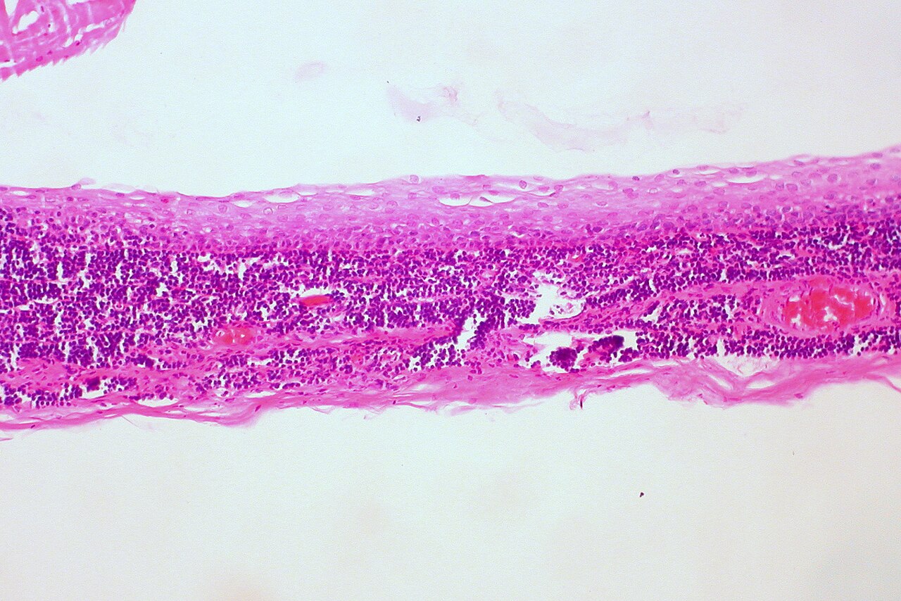
Branchial cleft cysts are significant as a common congenital neck mass in children, representing nearly 20% of pediatric neck masses. Their diagnosis is typically clinical due to their consistent location anterior to the sternocleidomastoid muscle, though in adults malignancy must be ruled out because carcinomas of the tonsil, tongue base, and thyroid can present similarly. The cyst wall is composed of squamous epithelium in 90% of cases, with lymphoid infiltrate. Treatment options include conservative management or surgical excision, though recurrence is common due to incomplete excision, as tracts often pass near important structures such as the internal jugular vein, carotid artery, or facial nerve. An alternative, less invasive treatment is ultrasound-guided sclerotherapy. The condition highlights the importance of understanding embryonic development and the potential for congenital anomalies to present later in life, often after an upper respiratory tract infection triggers infection of a previously unnoticed mass.
Did You Know?
- Second branchial cleft cysts represent 90 to 95% of all branchial cleft cysts.
- Infection of second branchial cleft cysts can compress the trachea, causing respiratory problems, or the esophagus, causing dysphagia.
Embryonic Origins and Tissue Architecture
These cysts are, at their core, vestiges of a developmental process shared with our fish ancestors. During early prenatal life, the human embryo forms four branchial clefts—structures homologous to the gill apparatus seen in aquatic vertebrates. In normal development, the first cleft matures into the external auditory canal while the remaining three are expected to obliterate entirely. When this process falters, typically involving the second, third, or fourth cleft, a persistent cavity remains. The most common mechanism is a failure of the second branchial arches and the epicardial ridge to fuse properly in the lower neck. The resulting cyst wall is not a random collection of cells; it is lined predominantly by squamous epithelium in roughly ninety percent of cases, though columnar cells, sometimes ciliated, or a blend of both epithelial types may be present. A distinctive feature is the dense lymphoid infiltrate surrounding the cyst, often with prominent germinal centers and a handful of subcapsular lymph sinuses, giving the lesion a histological signature that sets it apart from other neck swellings.
Clinical Presentation and the Diagnostic Dilemma
Most patients first notice a branchial cleft cyst in late childhood or the early adult years, when a previously silent, solitary, painless swelling in the upper neck—typically situated anterior to the sternocleidomastoid muscle—suddenly becomes symptomatic after an upper respiratory tract infection triggers inflammation. If a fistulous opening to the skin is present, it too remains quiet until infection sets in. In pediatric populations, these lesions account for nearly one-fifth of all neck masses, making them a common diagnosis in young patients. The clinical diagnosis is straightforward in many cases because of the lesion's predictable anatomical position. However, when a cystic neck mass appears in an adult, clinicians must treat it as a potential malignancy until evidence proves otherwise, since carcinomas of the tonsil, tongue base, and thyroid can all masquerade as cystic neck swellings. A useful distinguishing feature: unlike a thyroglossal duct cyst, a branchial cleft cyst does not elevate or descend with swallowing. Additionally, the discovery of bilateral skin pits should prompt evaluation for branchio-oto-renal syndrome.
Anatomical Classification and Regional Variations
Although all four embryonic clefts can give rise to persistent cysts, their clinical behavior and location vary dramatically. Second cleft cysts dominate the landscape, representing ninety to ninety-five percent of all branchial neck cysts. They characteristically sit anterior to the sternocleidomastoid, posterior to the submandibular gland, and lateral to the carotid sheath, with a skin pit sometimes visible in the anterior neck above the hyoid bone. First cleft cysts, by contrast, are periauricular in position, always found in or adjacent to the parotid gland, lateral to the facial nerve and running parallel to the external auditory canal; they make up about eight percent of neck sinuses and cysts. Third cleft cysts are rare and tend to appear in the posterior triangle of the upper neck or the anterior triangle of the lower neck. Fourth cleft abnormalities are not true cysts but rather sinus tracts or fistulae extending from the apex of the pyriform sinus down to the anterior lower neck, usually adjacent to the left thyroid lobe. Each type carries its own pattern of potential complications depending on the structures it abuts.
Treatment Challenges and Complication Risks
Management of branchial cleft cysts ranges from watchful waiting to definitive surgical excision, but neither path is without difficulty. When surgeons attempt complete excision, recurrence remains a significant concern, most often because the cyst's tract courses in close proximity to critical structures—the internal jugular vein, the carotid artery, and the facial nerve—making total removal impractical and carrying a high risk of iatrogenic injury. Infection adds another layer of complexity: a second cleft cyst that becomes inflamed can compress the trachea and produce respiratory compromise, press against the oesophagus and cause dysphagia, or irritate the sternocleidomastoid muscle and trigger torticollis. Fourth cleft anomalies, if they enlarge rapidly in a child, can similarly compress the trachea, and infection in this region may precipitate acute infectious thyroiditis. For patients in whom surgery poses unacceptable risk, ultrasound-guided sclerotherapy offers a less invasive alternative, collapsing the cyst cavity without the need to dissect through the dense web of vascular and neural anatomy that surrounds these lesions.
Gallery
Frequently Asked Questions
Who is Branchial cleft cyst?
It is a congenital swelling that sits in the upper neck, usually just in front of the sternocleidomastoid muscle, and it most often reveals itself in late childhood or early adulthood. Rather than being a tumor, it is a developmental remnant left behind during embryogenesis.
What are Branchial cleft cyst's powers/role?
Its signature move is presenting as a solitary, painless lump that can suddenly swell and become infected after a routine upper respiratory tract infection. In a subset of cases it also forms a small fistulous tract that opens onto the skin surface.
Why is Branchial cleft cyst important?
It makes up nearly 20% of all neck masses encountered in children, placing it among the most common congenital cervical lesions seen in pediatric and otolaryngology practice. Second branchial cleft cysts account for 90–95% of all cases, far outpacing third- and fourth-cleft variants.
What is Branchial cleft cyst's origin story?
It traces back to the early prenatal period, when the second, third, or fourth branchial cleft fails to fully obliterate or the second branchial arches and epicardial ridge fail to fuse as they should. That embryonic misstep leaves a small epithelial-lined sac that can later accumulate fluid and become a visible neck mass.
More in Genetic disorders with OMIM but no gene 1-24
Spotted an error? Know more?
Reader corrections go straight into our review queue. Suggest an edit · How this site is sourced