
Brain abscess
An abscess within brain tissue from infection or trauma.

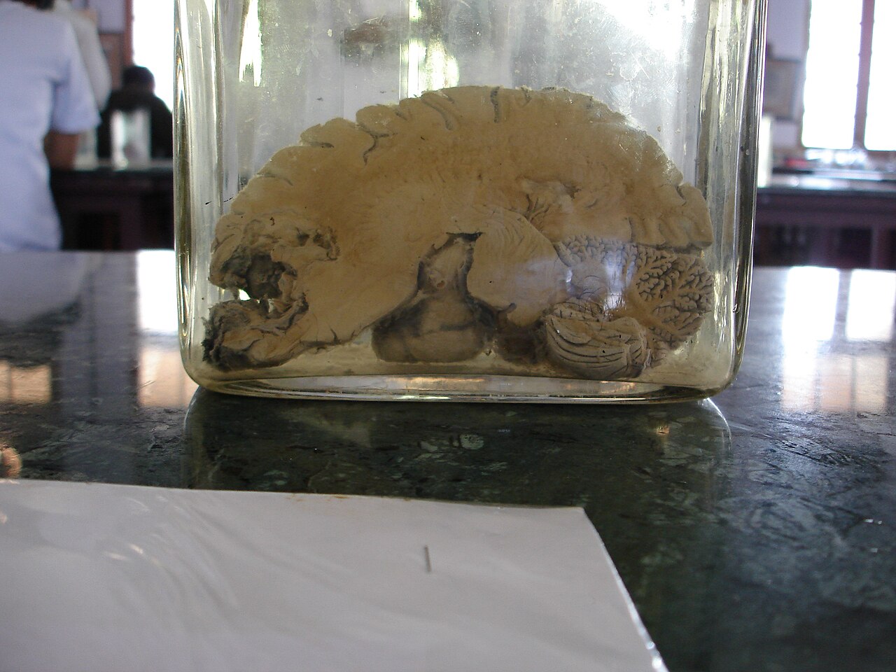
Netha Hussain · CC BY-SA 3.0
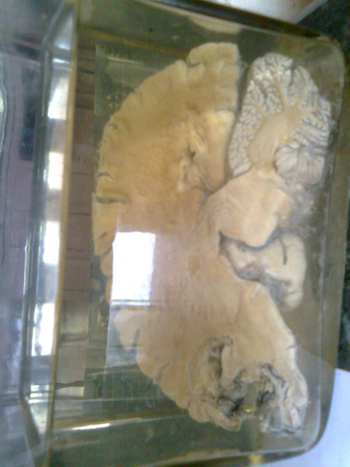
A brain abscess (or cerebral abscess) is an abscess within the brain tissue caused by inflammation and collection of infected material from local or remote infectious sources. It may also be introduced through a skull fracture following head trauma or surgical procedures. Brain abscess is usually associated with congenital heart disease in young children and can occur at any age, most frequently in the third decade of life.
- Field
- Infectious disease, neurosurgery
- Known for
- Ring-enhancing lesion on CT, triad of fever, headache, and focal neurologic findings
- Common organisms
- Staphylococcus aureus, streptococci, Bacteroides, Fusobacterium, Enterobacteriaceae
- Treatment
- Intravenous antibiotics, hyperbaric oxygen therapy, surgical drainage
- Prognosis
- Death in about 10% of cases; good outcome in about 70%
Lore & Background
Brain abscess is a space-occupying lesion that causes increased intracranial pressure, infection, and focal neurologic damage. The classic triad of fever, headache, and focal neurologic findings occurs in only 20% of people, but is highly suggestive when present. Headache is characteristically worse at night and in the morning due to natural increases in intracranial pressure when supine, and may be accompanied by morning vomiting.
Reader's Guide
Brain abscess remains a serious condition with a mortality rate of about 10%, though outcomes have improved since the 1960s due to better imaging, neurosurgery, and antibiotics. Diagnosis is established by CT with contrast, showing a ring-enhancing lesion after 4–5 days. Lumbar puncture is contraindicated because it may cause brain herniation. Treatment includes lowering intracranial pressure, intravenous antibiotics, hyperbaric oxygen therapy, and surgical drainage. The location of the abscess often suggests the primary infection source, such as middle ear infections leading to lesions in the middle and posterior cranial fossae. The Pennybacker system, pioneered by neurosurgeon Joseph Buford Pennybacker, introduced the use of pre-operative imaging and post-operative monitoring that remains standard today.
Did You Know?
- Lumbar puncture is contraindicated in brain abscess because it may cause brain herniation.
- Hyperbaric oxygen therapy reduces intracranial pressure and acts as a bactericide against anaerobic flora.
Origins and Routes of Infection
A brain abscess is almost never a spontaneous event confined to the brain itself. In the vast majority of cases, a primary infectious lesion exists elsewhere in the body, and the pathogen travels to the brain through local or distant routes. Local sources include middle-ear infections, dental abscesses, paranasal sinus disease, mastoid air-cell infections, and epidural abscesses. Distant sources can involve the lungs, heart, or kidneys. In young children, congenital heart disease—particularly with right-to-left shunts—serves as a well-recognized gateway, often seeding the territory supplied by the middle cerebral artery. Head trauma producing compound skull fractures, retained bullets, or surgical procedures can also introduce organisms directly into brain tissue. The anatomical position of the resulting abscess often acts as a map back to its origin: middle-ear infections tend to produce lesions in the middle and posterior cranial fossae, while frontal and ethmoid sinus disease more commonly yields collections near the subdural sinuses. Clinically, the condition can appear at any age but peaks during the third decade of life. Because failure to identify and treat the primary lesion leads to relapse, a thorough search for the source is considered essential.
Clinical Presentation and the Classic Triad
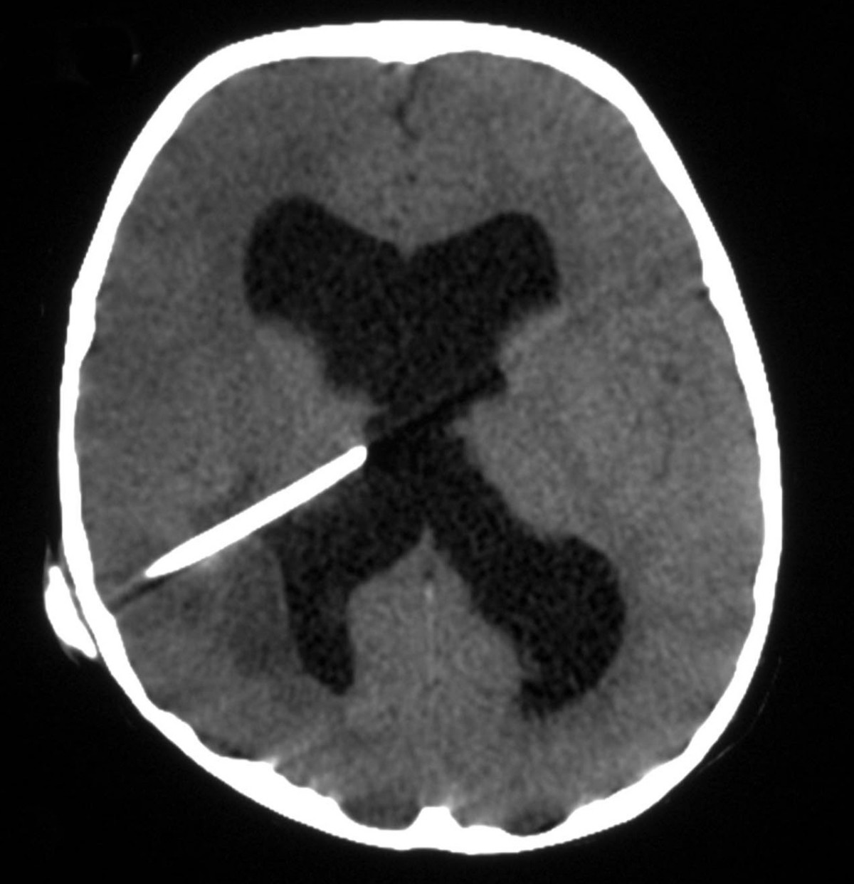
The textbook description of a brain abscess centers on a triad of fever, headache, and focal neurological deficits, yet this full constellation appears in only about one-fifth of affected individuals. More commonly, patients present with a rapidly progressive combination of headache, drowsiness, confusion, seizures, hemiparesis, or speech difficulties accompanied by fever. The headache carries a distinctive temporal pattern: it worsens at night and upon waking in the morning, a reflection of the natural rise in intracranial pressure when the patient lies supine. That same pressure elevation stimulates the medullary vomiting center and the area postrema, producing the characteristic morning vomiting. Beyond these shared features, symptoms shift according to the abscess's precise location. A cerebellar collection, for example, can compress the brain stem and precipitate hydrocephalus, adding a separate layer of neurological compromise. In occasional cases, a stiff neck may be detected on examination, which can misleadingly point toward meningitis rather than the true diagnosis. The underlying mechanism is a convergence of three processes: a space-occupying lesion raising intracranial pressure, active infection generating fever and fatigue, and direct destruction of focal brain tissue producing deficits such as hemiparesis or aphasia.
Microbiology and Predisposing Conditions
The microbial landscape of brain abscess is dominated by anaerobic and microaerophilic cocci alongside gram-negative and gram-positive anaerobic bacilli, and many infections are polymicrobial. The most frequently isolated organisms include Staphylococcus aureus, aerobic and anaerobic streptococci (with Streptococcus intermedius standing out), Bacteroides, Prevotella, and Fusobacterium species, as well as Enterobacteriaceae and Pseudomonas. Less commonly encountered are Haemophilus influenzae, Streptococcus pneumoniae, and Neisseria meningitidis. In immunocompromised patients, fungi and parasites become prominent culprits. The specific pathogen often mirrors the patient's underlying condition: sinus and dental infections favor streptococci, anaerobic gram-negative bacilli, and Fusobacterium; penetrating trauma brings S. aureus and Clostridium into play; pulmonary disease adds Actinomyces and Nocardia; congenital heart disease selects for aerobic and microaerophilic streptococci; HIV-associated abscesses implicate Toxoplasma gondii, Cryptococcus, and Listeria; transplant recipients face Aspergillus, Mucorales, and Candida; and neutropenic patients are vulnerable to aerobic gram-negative bacilli and mold infections. In AIDS patients, poliovirus, Toxoplasma, and Cryptococcus neoformans are the most frequently associated organisms, though the latter more often presents with meningitis rather than a discrete abscess.
Imaging, Diagnosis, and the Ring-Enhancing Lesion
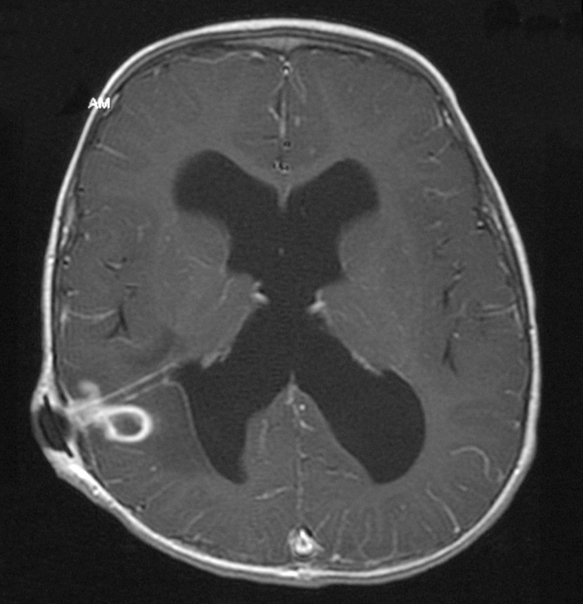
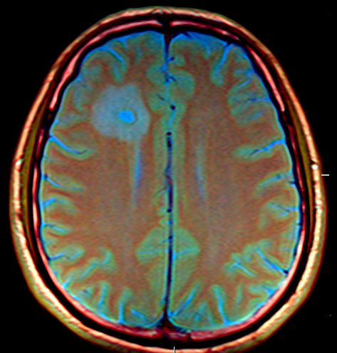
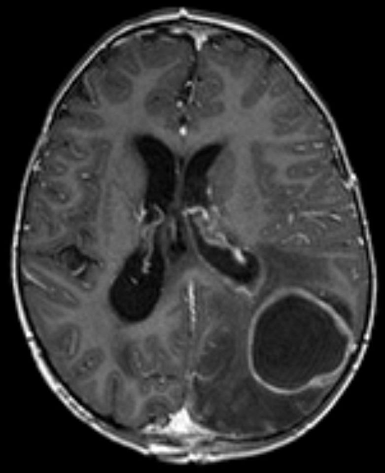
Computed tomography with intravenous contrast remains the cornerstone for establishing the diagnosis of brain abscess. In the earliest inflammatory stage, known as cerebritis, the lesion lacks a defined capsule, making it notoriously difficult to separate from other space-occupying lesions or cerebral infarcts on imaging. Within roughly four to five days, however, the surrounding inflammation and necrotic brain tissue become enclosed by a capsule. Because intravenously administered contrast material cannot penetrate this barrier, it pools around the periphery of the lesion, producing the classic ring-enhancing appearance against a relatively dark center. Importantly, this radiological sign is not pathognomonic: cerebral hemorrhages and certain brain tumors can generate a similar ring pattern. Clinical context—particularly a rapidly progressive course combined with fever, focal neurological deficits, and signs of elevated intracranial pressure—helps distinguish a true abscess from these mimics. A critical diagnostic caveat is that lumbar puncture, a routine procedure in many central nervous system infections, is strictly contraindicated here. Removing cerebrospinal fluid can disrupt intracranial pressure balances and precipitate dangerous brain herniation, a risk shared by all space-occupying intracranial lesions.
Gallery
Frequently Asked Questions
Who is Brain abscess?
Brain abscess is a localized pocket of pus that forms inside brain tissue when bacteria or other pathogens trigger a severe inflammatory response. It falls under the domains of infectious disease and neurosurgery, and although it can appear at any age, it most often strikes people in their twenties and thirties.
What are Brain abscess's powers/role?
Its signature move is producing a ring-enhancing lesion on CT imaging alongside a classic triad of fever, persistent headache, and focal neurological deficits. It is also a well-recognized trigger for seizures, and the usual organisms behind it include Staphylococcus aureus, various streptococci, Bacteroides, Fusobacterium, and Enterobacteriaceae.
Why is Brain abscess important?
It matters because it can arise from seemingly unrelated sources—congenital heart defects in young children, a skull fracture after head trauma, or post-surgical complications—making diagnosis genuinely tricky. Its association with seizures and the need to distinguish it from brain tumors or other ring-enhancing lesions keep it a critical entity in neurosurgical practice.
What's Brain abscess's origin story?
It usually begins when an infection from a nearby site (ear, sinuses, lungs) or a distant one (such as endocarditis in a child with a congenital heart defect) seeds bacteria into brain parenchyma. It can also be introduced directly through a fractured skull or during a neurosurgical procedure.
More in Disorders causing seizures 1-24
Spotted an error? Know more?
Reader corrections go straight into our review queue. Suggest an edit · How this site is sourced