
Anomalous left coronary artery from the pulmonary artery
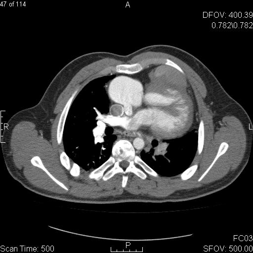
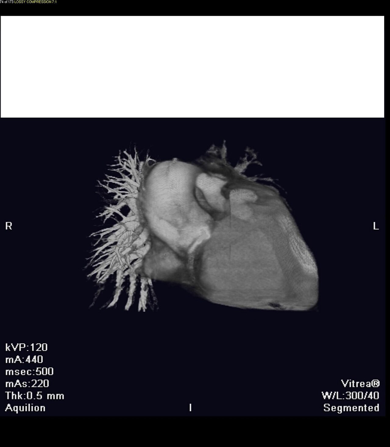
Rare congenital heart defect with left coronary artery from pulmonary artery.

Jto410 · CC BY-SA 3.0
Anomalous left coronary artery from the pulmonary artery (ALCAPA), also known as Bland-White-Garland syndrome or White-Garland syndrome, is a rare congenital anomaly in which the left coronary artery arises from the pulmonary artery instead of the aortic sinus. This condition occurs in approximately 1 in 300,000 liveborn children and accounts for between 0.24 and 0.46% of all congenital heart disease cases. Without treatment, about 90% of affected infants die within the first year, though rare cases of survival to adulthood have been documented.
- Field
- Congenital heart disease
- Known for
- Rare congenital anomaly where left coronary artery arises from pulmonary artery
- Incidence
- Approximately 1 in 300,000 liveborn children
- Percentage of congenital heart disease
- 0.24 to 0.46%
- Mortality without treatment
- Approximately 90% within first year
Lore & Background
In fetal life, high pressure in the pulmonary artery and fetal shunts allow oxygen-rich blood to flow through the anomalous left coronary artery. After birth, pulmonary artery pressure decreases, and the myocardium supplied by the left coronary artery becomes dependent on collateral blood flow from the right coronary artery. As pressure in the right coronary artery exceeds that in the left, collateral circulation increases, eventually leading to retrograde blood flow from the right coronary artery into the left coronary artery and then into the pulmonary artery, forming a left-to-right shunt.
Reader's Guide
The development of symptoms in ALCAPA depends heavily on the amount of collateral development. Infants with few collaterals experience myocardial ischemia, presenting with signs of heart failure such as dyspnea and tachypnea, or more subtle signs like crying during feeding, sweating, failure to thrive, and irritability. Patients with significant collateral circulation may survive to adulthood but remain at risk for chronic ischemia, sudden cardiac arrest, heart failure, or malignant arrhythmia. Diagnosis historically relied on conventional angiography, but today echocardiography is commonly used, along with computed tomography and magnetic resonance imaging. Surgery is indicated in all patients regardless of symptoms, with reimplantation of the left coronary artery into the aorta being the preferred approach. Reestablishment of a two-coronary system can make malignant arrhythmia disappear even in adults. Long-term follow-up is necessary to detect recurrent left ventricle dysfunction.
Did You Know?
- Approximately 90% of patients die within the first year if left untreated.
- Surgery is indicated in all patients with ALCAPA independent of symptoms.
Anatomical Journey and Variants
The left anterior descending artery takes its origin from the left main coronary artery, emerging just beyond the pulmonary artery. From that point it advances along the anterior interventricular sulcus, tracing this groove downward until it reaches the notch at the cardiac apex. A well-recognized variant, the wraparound LAD, does not stop at the apex; instead it curls around the tip of the heart and terminates on the posterior wall. Though exceedingly rare, the medical literature has also recorded instances in which the LAD arises from the right aortic sinus rather than its expected left-sided origin. These unusual courses, while uncommon, carry real clinical significance because they shift the distribution of myocardial territory that depends on the vessel. The typical path—descending the anterior interventricular groove—remains the overwhelming norm, yet the documented variants serve as a reminder that individual cardiac anatomy can depart meaningfully from the textbook picture.
Branching Architecture and Segmentation
The LAD gives rise to two principal categories of branches, each serving a distinct anatomical role. Septal perforators leave the main trunk at roughly right angles to the heart's surface, penetrating the myocardium to irrigate the anterior two-thirds of the interventricular septum. Diagonal branches, in contrast, stay on the epicardial surface, running along the lateral wall of the left ventricle and reaching the anterolateral papillary muscle. For angiographic and interventional purposes, the vessel is conventionally divided into three segments. The proximal segment spans from the LAD's origin to the first septal or diagonal branch, though definitions differ on which landmark marks the boundary. The middle segment continues from that point to roughly the midpoint of the remaining distance to the apex, or to the location where the artery forms a visible angle on a right anterior oblique angiographic view, often near the second diagonal. The distal segment covers the final stretch to the apex and, in wraparound variants, extends beyond it onto the posterior wall.
Myocardial Supply and Criticality
In terms of perfusion, the LAD is responsible for the anterior region of the left ventricle, a territory that includes the anterolateral myocardium, the apex, the anterior interventricular septum, and the anterolateral papillary muscle. In typical anatomy this single vessel delivers roughly 45 to 55 percent of the total arterial blood flow to the left ventricle, a share that makes it the most critical coronary artery for myocardial perfusion. No other branch of the coronary system matches the LAD in the sheer breadth of ventricular tissue it sustains. This outsized contribution explains why the vessel is consistently described as the most important artery supplying the left ventricle. The concentration of so much dependent myocardium on one channel means that any compromise to its lumen—whether from progressive atherosclerosis or acute thrombosis—carries consequences far greater than an equivalent blockage in a smaller branch. The LAD's dominance in ventricular supply is the anatomical foundation for the clinical urgency that surrounds any threat to its patency.
The Widow-Maker and Clinical Diagnosis
Atherosclerotic disease within the LAD can progress through plaque rupture to complete arterial occlusion, triggering a myocardial infarction. When the proximal segment is the site of blockage, the event is colloquially called the widow-maker heart attack, a name that reflects the poor prognosis that typically follows. The severity of this particular occlusion is amplified by the enormous myocardial territory the LAD supplies, and the danger escalates further in patients whose vessel follows an anomalous or wraparound course, because additional tissue on the posterior wall becomes vulnerable to ischemia. Among all coronary occlusions, proximal LAD blockage stands out for its lethality and the breadth of tissue at risk. In the acute setting, a standard 12-lead electrocardiogram remains the best diagnostic tool for identifying the electrical signature of this infarction. The combination of the LAD's critical supply role, the potential for sudden and complete occlusion, and the availability of rapid ECG-based diagnosis makes this vessel a central focus of both preventive cardiology and emergency interventional practice.
Gallery
Frequently Asked Questions
Who is Anomalous left coronary artery from the pulmonary artery?
ALCAPA, also called Bland-White-Garland syndrome, is a congenital heart defect present at birth in which the left coronary artery connects to the pulmonary artery rather than to the aortic sinus. It is classified as a rare anomaly within the broader family of congenital heart diseases.
What is ALCAPA's mechanical 'power' or role in the body?
In a typical heart the left coronary artery delivers oxygen-rich blood to the myocardium, but in ALCAPA it instead draws blood from the lower-pressure pulmonary artery. The result is that the heart muscle is starved of properly oxygenated fuel, impairing its ability to pump effectively.
Why is ALCAPA considered important in the canon of congenital heart disease?
Although it accounts for only about 0.24 to 0.46 percent of all congenital heart defects, its near-certain fatality without intervention makes it one of the most urgent diagnoses in pediatric cardiology. Early surgical correction is the single factor that transforms the prognosis from almost universally fatal to survivable.
How rare is ALCAPA's 'appearance' in the general population?
The anomaly is detected in roughly one out of every 300,000 liveborn children, placing it among the rarer congenital cardiac conditions. Its low incidence means that each new diagnosis represents a significant and uncommon event in a child's medical history.
More in Syndromes 1-24
Spotted an error? Know more?
Reader corrections go straight into our review queue. Suggest an edit · How this site is sourced