Optical microscope
Uses visible light and lenses to magnify small objects.

Unknown author · Public domain
An optical microscope, also called a light microscope, uses visible light and a set of lenses to produce enlarged images of tiny objects. It is the oldest kind of microscope; the compound version we know today first appeared in the 1600s. Basic models can be quite simple, but many complex designs exist to improve resolution and contrast.
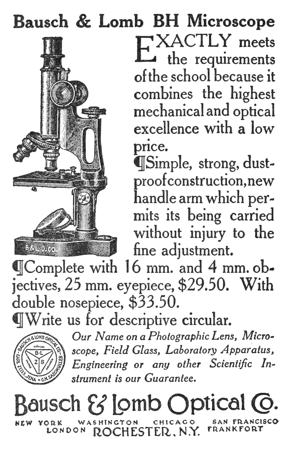
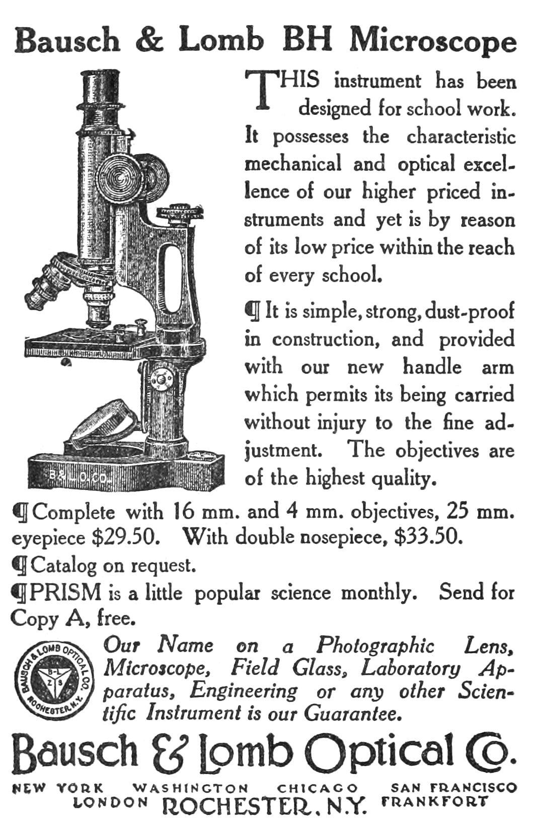
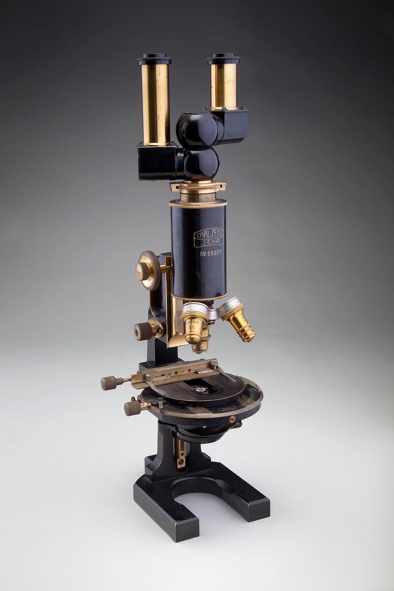
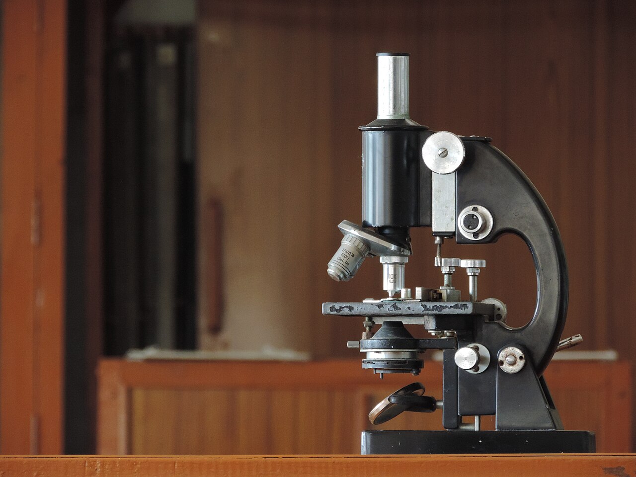
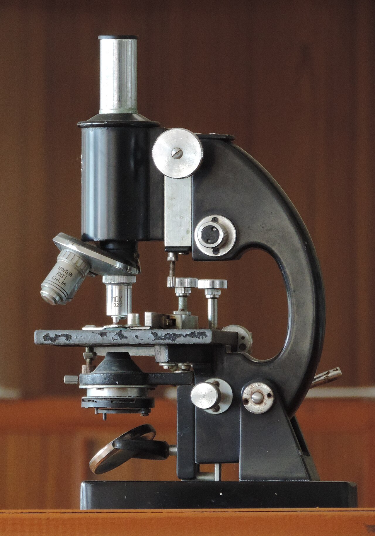
Samples sit on a stage and can be viewed directly through one or two eyepieces. A rotating turret between the stage and the eyepiece usually holds several objective lenses with different magnifications, letting the user adjust the zoom. In high-power microscopes, both eyepieces show the same image, while a stereo microscope shows slightly different images to create a 3-D effect. A camera can also capture the magnified picture, called a micrograph.
Lighting methods vary. Transparent objects can be lit from below; solid objects can be lit with light passing through the objective lens (bright field) or around it (dark field). Polarized light helps determine crystal orientation in metals. Phase-contrast imaging boosts contrast by highlighting small differences in refractive index.
The best resolving power of optical microscopes is usually around 200 nanometers, limited by the diffraction of visible light. Higher magnifications are possible but don't reveal new details. Alternatives that don't use visible light include scanning electron microscopy, transmission electron microscopy, and scanning probe microscopy, which can achieve much greater magnifications.
**Types**
There are two basic types: simple and compound. A simple microscope uses a single lens or group of lenses for magnification. A compound microscope uses one set of lenses to enlarge the image produced by another set, achieving much higher magnification. Most modern research microscopes are compound; some cheaper digital microscopes are simple single-lens designs. Compound microscopes can be further divided into many types, differing in optics, cost, and purpose.
**Simple microscope**
A simple microscope uses a lens or set of lenses to enlarge an object through angular magnification alone, giving the viewer an upright, enlarged virtual image. This is found in magnifying glasses, loupes, and eyepieces for telescopes and microscopes.
**Compound microscope**
A compound microscope uses a lens close to the object (the objective) to collect light and focus a real image inside the microscope. That image is then magnified by another lens or group of lenses (the eyepiece), giving the viewer an enlarged, inverted virtual image. This objective-eyepiece combination allows much higher angular magnification. For an object of height *h*, it can at most occupy an unmagnified angular size θ₀ = *h*/*d₀* while remaining in focus, achieved when placed at the near point distance *d₀* of the eye (about 11 cm). The virtual image created by the compound microscope achieves an angular size θ = –*hs* / (*f_objective* *f_eyepiece*), where *s* is the distance between the neighboring objective and eyepiece focal points. This gives an angular magnification of θ/θ₀ = –*sd₀* / (*f_objective* *f_eyepiece*). Common compound microscopes often have exchangeable objective lenses for quick magnification changes, and they also enable advanced illumination setups like phase contrast.
**Other microscope variants**
Many compound microscope designs exist for specialized purposes, differing in physical layout:
- **Stereo microscope:** A low-powered microscope providing a stereoscopic view, often used for dissection. - **Comparison microscope:** Has two separate light paths, allowing direct comparison of two samples, one image per eye. - **Inverted microscope:** Studies samples from below; useful for cell cultures in liquid or metallography. - **Fiber optic connector inspection microscope:** Designed for inspecting connector end-faces. - **Traveling microscope:** For studying samples requiring high optical resolution.
- type
- Scientific instrument
- first_appearance
- 17th century (compound form)
- maximum_resolving_power
- Around 200 nanometers
- basic_types
- Simple and compound
- common_illumination_methods
- Bright field, dark field, polarized light, phase-contrast
Lore & Background
The optical microscope, also referred to as a light microscope, is a type of microscope that commonly uses visible light and a system of lenses to generate magnified images of small objects. Optical microscopes are the oldest type of microscope, with the present compound form first appearing in the 17th century. Basic optical microscopes can be very simple, although many complex designs aim to improve resolution and sample contrast. Objects are placed on a stage and may be directly viewed through one or two eyepieces on the microscope. A range of objective lenses with different magnifications are usually mounted on a rotating turret between the stage and eyepiece(s), allowing magnification to be adjusted as needed. In high-power microscopes, both eyepieces typically show the same image, but with a stereo microscope, slightly different images are used to create a 3-D effect. A camera may be used to capture the magnified image (micrograph). Objects can be lit in a variety of ways. Transparent objects can be lit from below and solid objects can be lit with light coming through (bright field) or around (dark field) the objective lens. Polarised light may be used to determine crystal orientation of metallic objects. Phase-contrast imaging can be used to increase image contrast by highlighting small details of differing refractive index.
Reader's Guide
The optical microscope is a foundational instrument in biology, materials science, and medicine, providing the first detailed views of cells, microorganisms, and small structures. Its maximum resolving power is typically limited to around 200 nanometers due to the diffraction limit of visible light, meaning larger magnifications beyond this point do not reveal additional detail. Despite this limitation, optical microscopes remain widely used because they can observe living specimens in natural color and are relatively simple and affordable. The development of specialized variants, such as the phase-contrast microscope, polarizing microscope, and confocal microscope, has expanded its utility for specific applications like examining transparent samples, crystalline materials, and fluorescent markers. Alternatives that do not use visible light, such as scanning electron microscopy and transmission electron microscopy, can achieve much greater magnifications but require different sample preparation and cannot image living organisms. The digital microscope, which incorporates a camera and computer control, has further enhanced analysis capabilities, allowing measurements and quantitation of images. Overall, the optical microscope remains an essential tool for direct visual observation and initial investigation across many scientific disciplines.
Did You Know?
- The compound form of the optical microscope first appeared in the 17th century.
- The maximum resolving power of optical microscopes is typically limited to around 200 nanometers due to the diffraction limit of visible light.
- A stereo microscope uses slightly different images in each eyepiece to create a 3-D effect.
- Phase-contrast imaging can increase image contrast by highlighting small details of differing refractive index.
Historical Roots and Design Evolution
The optical microscope holds the distinction of being the earliest form of magnification instrument ever developed. While basic single-lens devices have ancient roots, the compound configuration that dominates modern laboratories first emerged during the 17th century. Over the centuries, the fundamental architecture has remained remarkably consistent: a specimen rests on a stage, light passes through or around the sample, and a series of lenses enlarges the resulting image for the observer. Yet the range of complexity is vast. At one end sit rudimentary single-lens magnifiers, essentially a convex lens producing an upright virtual image, while at the other end are elaborate research instruments incorporating multiple lens groups, precision illumination systems, and specialized optical filters. The compound design, which pairs an objective lens near the specimen with a separate eyepiece system, became the standard for virtually all serious scientific work, offering far greater angular magnification than any single-element device could achieve. This evolutionary arc, from a simple glass element to a multi-lens research platform, underscores how a single optical principle has been refined across four centuries of ingenuity.
The Compound Lens System and Magnification
The core innovation of the compound microscope lies in its two-stage magnification architecture. An objective lens positioned close to the specimen collects light and projects a real, inverted image inside the body tube. A second optical group, the eyepiece, then takes that intermediate image and enlarges it further, delivering to the viewer an inverted virtual image of substantially greater angular size. The theoretical angular magnification depends on the separation between the focal points of the two lens groups and the focal lengths of each, meaning that shorter focal lengths in both the objective and eyepiece yield greater enlargement. In practice, most compound microscopes mount a rotating turret of interchangeable objective lenses between the stage and the eyepiece, letting the user swap magnification levels with a quarter-turn. This modular approach, combined with the ability to stack advanced illumination methods such as phase contrast, makes the compound design the workhorse of biological and materials research. By contrast, the simpler single-lens microscope, found in inexpensive digital devices and basic magnifying glasses, relies on angular magnification from one element alone and cannot match the resolving capability of its compound counterpart.
Illumination Strategies and Specialized Variants
How light reaches and passes through a specimen is one of the most consequential design choices in optical microscopy. Transparent samples are commonly illuminated from beneath the stage, while opaque or solid objects may be lit either directly through the objective in bright-field mode or with light directed around the objective axis in dark-field mode. Polarized light introduces an additional analytical dimension, enabling the determination of crystal orientation in metallic specimens. Phase-contrast illumination, a particularly elegant technique, amplifies subtle differences in refractive index so that otherwise invisible internal structures become visible. These illumination methods have spawned a rich family of specialized instruments. A petrographic microscope incorporates a polarizing filter, a rotating stage, and a gypsum plate to study mineral crystals whose optical properties shift with orientation. An inverted microscope flips the geometry so the objective sits below the stage, making it ideal for observing living cell cultures in liquid or for metallographic work. A stereo microscope feeds slightly different images to each eye to produce a three-dimensional view, a feature especially valued in dissection and assembly tasks. A comparison microscope splits the light path entirely, presenting two separate samples side by side in a single binocular view. Each variant represents a tailored solution to a specific investigative need.
The Diffraction Barrier and Beyond
No matter how sophisticated the lens train or how elaborate the illumination scheme, the optical microscope confronts a hard physical ceiling. Because visible light waves diffract at apertures, the smallest resolvable feature is capped at roughly 200 nanometers. Pushing the magnification higher beyond this threshold produces a larger image but no additional structural detail; the extra enlargement is, in effect, empty. This diffraction limit is not an engineering shortcoming but a consequence of the wavelength of the light itself. For researchers who need to probe structures smaller than this boundary, the field has turned to entirely different physical principles. Scanning electron microscopy and transmission electron microscopy replace photons with beams of electrons, whose wavelengths are orders of magnitude shorter, thereby achieving far greater resolving power. Scanning probe microscopy takes a different route still, using a physical tip to raster across a surface and map topography at the atomic scale. These techniques do not compete with the optical microscope so much as extend the observational frontier into regimes where visible light simply cannot reach.
Gallery
Frequently Asked Questions
Who is Optical microscope?
It's the founding member of the microscope family, with its compound design first showing up in the 17th century. Fans of the series treat it as the OG character every later magnification tool builds on.
What are Optical microscope's powers/role?
It channels visible light through a stack of glass lenses to inflate tiny specimens into visible detail, resolving structures down to roughly 200 nanometers. Its toolkit includes bright-field, dark-field, polarized-light, and phase-contrast illumination modes for different specimen types.
How does Optical microscope's story end?
There's no final episode because it's still a daily workhorse in biology labs and hospital pathology rooms. Its lasting legacy is simply that it opened the door to seeing the microscopic world at all.
Why is Optical microscope important?
It remains the most accessible and widely used instrument for examining cells, tissues, and microorganisms that the naked eye can't resolve. Both science and medicine depend on it as a first-line observation tool.
What's the difference between Optical microscope's two basic types?
The simple variant relies on a single lens for modest magnification, while the compound variant chains multiple lens groups together for far greater power. Both share the same visible-light principle but differ in how many optical stages the light passes through.
More in Science & Measurement 1-16
Spotted an error? Know more?
This is a living reference — every entry is fact-audited, and reader corrections feed straight into our audit queue. Suggest an edit · See this site's audit record