Acromesomelic dysplasia
Rare skeletal disorder causing shortened limbs and abnormal bone growth.

Acromesomelic dysplasia (AMD) is a rare skeletal condition marked by faulty bone and cartilage development. This results in shortened forearms, lower legs, hands, feet, fingers, and toes. The disorder usually becomes noticeable in early childhood. It is caused by mutations in one of several genes, with the main types linked to three key genes (NPR2, GDF5, BMPR1B), and is inherited in an autosomal recessive pattern, meaning a child must receive two copies of the abnormal gene—one from each parent—to develop the condition.
Signs and symptoms include inhibited growth of certain long bones, so the forearms, lower legs, hands, and feet do not grow in proportion to the rest of the body. Over time, individuals may have difficulty fully extending their arms or rotating them inward and outward. The bones in the fingers, toes, hands, and feet can become shorter and broader. During early childhood, progressive abnormal curvature of the spine may occur. Common symptoms are short stature, progressive degeneration, stiffness, tenderness, osteoarthritis, and abnormal bone shape. Infants with AMD typically have a normal birth weight but may show characteristic facial features such as macrocephaly, frontal bossing, occipital prominence, a slightly flattened midface, and an abnormally small, pug nose.
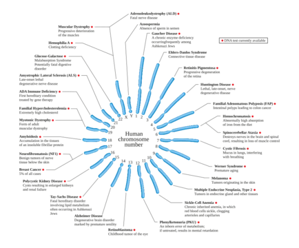
The condition is extremely rare. Genetic studies have identified specific mutations: AMD Maroteaux involves a mutation on chromosome 9p13-12, affecting the gene for natriuretic peptide receptor B (NPR2), a receptor for a hormone essential for bone growth. AMD Grebe dysplasia involves a mutation on chromosome 20q11.2, affecting the gene for growth and development factor-5 (GDF5). AMD with genital anomalies involves a mutation on chromosome 4q23-24, affecting a gene for a bone morphogenetic protein receptor, type 1B (BMPR1B). Genetic diseases are determined by the combination of genes received from both parents.
Diagnosis is based on clinical presentation, molecular analysis, electropherogram, and radiograph, typically made within the first few years of life. Key characteristics are identified, and molecular analysis examines the DNA of the affected person and their parents from a blood sample. Radiographs show abnormal growth plates and misshapen bones in the limbs, confirming abnormal development and premature fusion where the diaphyses of certain long bones meet their epiphy
- types
- 4 main types (Maroteaux, Grebe dysplasia, Du Pan syndrome, Acromesomelic dysplasia with genital anomalies)
- earliest_known_case
- Not specifically documented in standard medical literature
Lore & Background
Acromesomelic dysplasia (AMD) is an extremely rare genetic disorder that inhibits the growth of certain long bones, particularly the forearms and lower legs. The disease becomes apparent during early childhood, with infants typically having normal birth weight but later developing disproportionate growth. Over time, individuals may experience difficulty extending their arms or rotating them, and the bones in the fingers, toes, hands, and feet become shorter and broader. Progressive abnormal curvature of the spine, such as kyphosis and lumbar hyperlordosis, often develops during early childhood.
Reader's Guide
The significance of acromesomelic dysplasia lies in its demonstration of how specific genetic mutations disrupt bone and cartilage development. Five distinct types have been identified, each linked to a different gene mutation: Maroteaux type (chromosome 9p13-12, affecting natriuretic peptide receptor B), Grebe dysplasia (chromosome 20q11.2, affecting growth and development factor-5), and AMD with genital anomalies (chromosome 4q23-24, affecting bone morphogenetic protein receptor type 1B). Treatment is individualized and primarily symptomatic, including physical therapy, braces, corrective surgery for spinal curvature, and recombinant human growth hormone, which may increase growth velocity but does not cure the disease. The condition has a normal life expectancy but no chance of full recovery, with long-term effects including joint pain, arthritis, and short stature. With fewer than 100 reported cases worldwide, AMD is a model for understanding rare skeletal dysplasias and the role of specific genes in bone development.
Did You Know?
- AMD is caused by mutations in the NPR2, GDF5, or BMPR1B genes, each linked to a specific subtype of the disorder.
- Because AMD is inherited in an autosomal recessive pattern, both parents must carry a copy of the mutated gene for a child to be affected.
Clinical Presentation and Daily Living
Acromesomelic dysplasia first reveals itself during the earliest years of life, when parents notice that a child's forearms, lower legs, hands, and feet are failing to keep pace with the rest of the body. Although infants typically arrive at a normal birth weight, the disproportion becomes increasingly visible as growth proceeds. Over time, the small bones of the fingers, toes, hands, and feet grow shorter and broader, while the long bones of the forearms and lower legs remain stunted. This combination creates practical difficulties: fully straightening the arms, turning the palms downward, or rotating the hands outward all become physically challenging. In early childhood, the spine may begin to curve abnormally, developing progressive kyphosis and lumbar hyperlordosis. A constellation of secondary symptoms follows, including generalized stiffness, joint tenderness, and eventually osteoarthritis. The facial profile often carries distinctive markers—macrocephaly, a prominent forehead, a flattened midface, and a small, upturned nose—that help clinicians recognize the condition even before radiographic confirmation.
Genetic Architecture and the Five Subtypes
The disorder is passed down as an autosomal recessive trait, meaning a child must inherit two faulty copies of the responsible gene—one from each parent or through a de novo mutation—to develop the condition. Five distinct mutations have been identified, each producing a recognizable subtype. The Maroteaux form, linked to a mutation on chromosome 9p13-12, disrupts the natriuretic peptide receptor B protein, which normally relays signals from C-type natriuretic peptide, a hormone critical to bone elongation; affected individuals typically remain below 120 centimeters in height. Grebe dysplasia maps to chromosome 20q11.2 and involves growth and development factor-5, producing extreme joint malformations in the hands and feet with markedly reduced articulation, yet sparing overall stature. The genital-anomaly variant sits on chromosome 4q23-24 and impairs a bone morphogenetic protein receptor type 1B, adding congenital malformations of the reproductive tract. Osebold-Remondini and Du Pan syndrome round out the spectrum, the former featuring fused phalanges and carpal-tarsal coalitions, the latter characterized by underdeveloped fibulae, hands, and feet.
The Diagnostic Pathway
Because the condition is exceedingly rare, reaching a confident diagnosis requires a layered approach that typically unfolds within the first few years of life. Clinicians begin with a thorough physical examination and a detailed family history; a parent who carries the relevant chromosome often appears shorter than average, a subtle clue that can guide genetic counseling. Radiographs of the limbs then reveal the hallmark structural abnormalities—misshapen bones, irregular growth plates, and premature fusion where the shafts of long bones meet their ends. Molecular analysis adds a genetic dimension: a blood sample is drawn from the affected child and both parents, the DNA is extracted using an automated instrument, and the resulting sequence is checked for the specific chromosomal mutation. An electropherogram provides genotyping data that can be compared against a reference sequence and against relatives' profiles, confirming whether the variant was inherited or arose spontaneously. Together, these three pillars—clinical observation, imaging, and molecular genetics—allow the treating team to pinpoint which of the five subtypes is present and to tailor the long-term management plan accordingly.
Management and the Limits of Cure
No therapy currently eliminates the underlying genetic defect, so management is inherently palliative and tailored to each individual's subtype and symptom profile. Physical therapy occupies a central role and is most effective when begun in early childhood; targeted exercises, bracing, and casting can slow the progression of spinal curvature, while severe kyphosis or lumbar hyperlordosis may ultimately require corrective surgery. Recombinant human growth hormone, a synthetic version of the protein naturally secreted by the pituitary gland, represents another pillar of treatment. Administered over the long term, it does not cure the dysplasia but can modestly augment both muscle and skeletal growth in children and adolescents. Published case data report first-year growth velocities in the range of 3.6 to 4.2 centimeters per year, with one patient gaining 7.0 centimeters within twelve months; over a five-year course, height improved from roughly 1.2 to 1.8 standard deviations above the baseline. Because outcomes vary widely from person to person, the treating team must continually reassess which combination of interventions—exercise, orthotics, hormonal support, or surgical correction—best serves the individual at each stage of development.
Frequently Asked Questions
What is Acromesomelic dysplasia?
It is a rare skeletal disorder in which bone and cartilage fail to develop properly, producing noticeably shortened forearms, lower legs, hands, feet, fingers, and toes. The condition typically becomes apparent during early childhood.
What causes Acromesomelic dysplasia?
The disorder stems from mutations in one of several genes—most commonly NPR2, GDF5, or BMPR1B—and follows an autosomal recessive inheritance pattern. A child must inherit two defective copies, one from each parent, for the condition to manifest.
What are the main types of Acromesomelic dysplasia?
Four principal variants are recognized: Maroteaux type, Grebe dysplasia, Du Pan syndrome, and Acromesomelic dysplasia with genital anomalies. Each subtype carries its own specific pattern of skeletal involvement.
How does Acromesomelic dysplasia affect the body?
Growth of certain long bones is stunted, leaving the forearms, lower legs, hands, feet, fingers, and toes shorter than expected. The trunk and upper arms are generally less affected, giving a characteristic proportioning pattern.
Why is Acromesomelic dysplasia significant in genetics?
It serves as a clear example of how autosomal recessive inheritance and specific gene mutations can disrupt the intricate signaling pathways that govern bone and cartilage formation. Understanding its four distinct subtypes helps researchers map the roles of NPR2, GDF5, and BMPR1B in skeletal development.
More in Genetic diseases and disorders 1-24
Spotted an error? Know more?
This is a living reference — every entry is fact-audited, and reader corrections feed straight into our audit queue. Suggest an edit · See this site's audit record