
Respiratory failure
Respiratory failure impairs blood oxygen or carbon dioxide levels.

State of Hawaii · Public domain
Respiratory failure happens when the respiratory system can’t exchange gases properly, so the body can’t keep arterial oxygen, carbon dioxide, or both at normal levels. Low oxygen in the blood is called hypoxemia, while high carbon dioxide is hypercapnia. The condition is grouped into Type 1 or Type 2, depending on whether carbon dioxide is elevated, and it may be acute or chronic. In clinical studies, respiratory failure is typically defined by a faster breathing rate, abnormal blood gases (hypoxemia, hypercapnia, or both), and signs that breathing is harder than usual. Because the brain doesn’t get enough oxygen, it can lead to an altered state of consciousness. Normal reference values for partial pressure are oxygen (PaO₂) above 80 mmHg (11 kPa) and carbon dioxide (PaCO₂) below 45 mmHg (6.0 kPa).
**Cause** Many conditions can lead to respiratory failure, and the reasons differ depending on the type. Causes fall into several broad categories. Some reduce airflow into and out of the lungs—for example, a physical blockage from a foreign object or mass, or slowed breathing due to drugs or chest wall changes. Others impair the lungs’ blood supply, such as thromboembolic disorders or conditions that lower right heart output, like right heart failure and certain heart attacks. Still others limit the lung tissue’s ability to exchange oxygen and carbon dioxide between blood and air; any disease that damages lung tissue can do this. The most common causes (in no particular order) include infections, interstitial lung disease, and pulmonary edema.
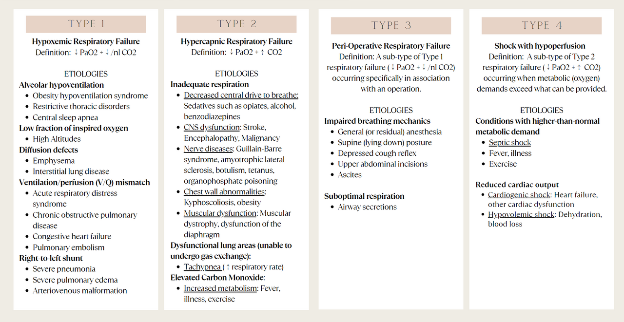
**Types** Respiratory failure is generally divided into four types, each with distinct features and main causes.
**Type 1** Type 1 respiratory failure involves low blood oxygen (hypoxemia), with PaO₂ below 60 mmHg, while carbon dioxide (PaCO₂) stays normal (normocapnia) or low (hypocapnia). The core problem is a failure of oxygenation. This type is caused by conditions that affect oxygenation, leading to lower-than-normal oxygen in the blood. These include low ambient oxygen (for instance, at high altitude); ventilation-perfusion mismatch (parts of the lung get oxygen but not enough blood to absorb it, as in pulmonary embolism, ARDS, COPD, or congestive heart failure); alveolar hypoventilation (reduced minute volume from weak respiratory muscles, as in acute neuromuscular disease—this can also cause Type 2 if severe); diffusion problems (oxygen can’t enter capillaries due to parenchymal disease, like pneumonia or ARDS); and right-to-left shunt (oxygenated blood mixes with deoxygenated venous blood, as in arteriovenous malformation, complete atelectasis, severe pneumonia, or severe pulmonary edema).
**Type 2** Type 2 respiratory failure shows hypoxemia (PaO₂ below 8 kPa or normal) along with hypercapnia (PaCO₂ above 6.0 kPa). The basic defect is inadequate alveolar ventilation, affecting both oxygen and carbon dioxide. It is defined by a buildup of carbon dioxide that the body produces but cannot eliminate. Underlying causes include increased airway resistance (COPD, asthma, suffocation); reduced breathing effort (from drugs, brain stem lesions, or extreme obesity); a decrease in the lung area available for gas exchange (as in chronic bronchitis); neuromuscular problems (Guillain-Barré syndrome, motor neuron disease); and chest wall deformities (kyphoscoliosis, ankylosing spondylitis, or flail chest).
**Type 3** Type 3 respiratory failure is a subtype of Type 1, with low PaO₂ (hypoxemia) and normal or low PaCO₂. It gets its own category because it is so common. Often called peri-operative respiratory failure, it is specifically linked to an operation, procedure, or surgery. The underlying problem usually involves lung atelectasis—a collapse of the functional gas-exchange units. Because atelectasis happens so often around the time of surgery, this form is also known as perioperative respiratory failure. After general anesthesia, a drop in functional residual capacity leads to collapse of dependent lung units.
**Type 4** Type 4 respiratory failure occurs when the body’s metabolic (oxygen) demands exceed what the cardiopulmonary system can supply. It often stems from hypoperfusion of the respiratory muscles, as seen in shock—for example, cardiogenic shock or hypovolemic shock. Patients in shock frequently have respiratory distress due to pulmonary edema (such as in cardiogenic shock). Lactic acidosis and anemia can also trigger Type 4. Still, Type 1 and Type 2 are the most widely accepted categories.
**Physical exam** On physical exam, patients with respiratory failure often show signs of impaired oxygenation (low blood oxygen). These include using accessory muscles to breathe or other signs of respiratory distress; altered mental status (like confusion or lethargy); clubbing of the fingertips; peripheral cyanosis (a bluish color on mucous membranes, fingers, or toes); tachypnea (faster breathing rate); and pale conjunctivae.
- definition
- Inadequate gas exchange causing abnormal arterial oxygen or carbon dioxide levels
- types
- Type 1, Type 2, Type 3, Type 4
- key_measurements
- PaO2 > 80 mmHg (11 kPa), PaCO2 < 45 mmHg (6.0 kPa)
- common_causes
- Infections, interstitial lung disease, pulmonary edema, COPD, asthma, neuromuscular disorders
- diagnostic_gold_standard
- Arterial blood gas (ABG) assessment
- prognosis
- One of three hospitalized cases is fatal
Lore & Background
Respiratory failure is organized into four types. Type 1 is characterized by hypoxemia with normal or low carbon dioxide, often due to ventilation-perfusion mismatch, diffusion problems, or shunts. Type 2 involves hypercapnia alongside hypoxemia, caused by inadequate alveolar ventilation from conditions like COPD, neuromuscular problems, or reduced breathing effort. Type 3 is a peri-operative form of Type 1 associated with lung atelectasis after surgery. Type 4 results from metabolic demands exceeding cardiopulmonary capacity, as in shock states.
Reader's Guide
Respiratory failure is a critical condition with significant clinical importance. Its classification into types guides treatment: Type 1 often requires oxygen therapy, while Type 2 typically needs non-invasive ventilation. The condition's high mortality—one in three hospitalized cases is fatal—underscores the need for prompt diagnosis via arterial blood gas and appropriate management of underlying causes. Understanding its etiologies, from infections to neuromuscular diseases, helps clinicians tailor interventions such as bronchodilators, antibiotics, or mechanical ventilation. The inclusion of peri-operative and shock-related types highlights its relevance across medical settings.
Did You Know?
- Respiratory failure causes an altered state of consciousness due to ischemia in the brain.
- Type 3 respiratory failure is specifically associated with operations or surgery and is often called peri-operative respiratory failure.
- The gold standard diagnostic test for respiratory failure is arterial blood gas (ABG) assessment.
- One of three hospitalized cases of acute respiratory failure is fatal.
Classification & Typology
Respiratory failure is fundamentally a breakdown in the lungs' ability to maintain normal arterial oxygen and carbon dioxide levels. Clinicians categorize this condition along two axes: whether carbon dioxide accumulates (distinguishing Type 1 from Type 2) and whether the onset is acute or chronic. Normal reference values place arterial oxygen partial pressure above 80 mmHg and carbon dioxide below 45 mmHg; deviation from these thresholds signals failure. Type 1 failure presents with dangerously low oxygen (below 60 mmHg) while carbon dioxide remains normal or even low. Type 2 failure adds a dangerous rise in carbon dioxide above 6.0 kPa, reflecting the body's inability to exhale the CO2 it has generated. Type 3, though mechanistically a subset of Type 1, has earned its own label because it is specifically tied to the surgical and perioperative setting. Type 4 describes a scenario where the body's metabolic oxygen demands outstrip what the cardiopulmonary system can deliver. In research settings, a formal diagnosis typically requires an elevated respiratory rate, abnormal blood gas values, and observable increased work of breathing.
Etiology & Pathophysiology
The roots of respiratory failure span three broad mechanistic categories. First, conditions that physically restrict airflow into and out of the lungs—whether through foreign-body obstruction, tumor masses, drug-induced suppression of breathing, or structural changes to the chest wall. Second, disorders that compromise the pulmonary blood supply, including thromboembolic events, right-sided heart failure, and certain myocardial infarctions that reduce cardiac output. Third, diseases that damage the lung parenchyma itself, impairing the tissue's capacity to swap oxygen and carbon dioxide between alveoli and capillaries; infections, interstitial lung disease, and pulmonary edema are among the most frequent culprits. Within Type 1 failure specifically, mechanisms include ventilation-perfusion mismatch (pulmonary embolism, ARDS, COPD, congestive heart failure), diffusion impairment from parenchymal damage, right-to-left shunting (arteriovenous malformations, complete atelectasis, severe edema), and low ambient oxygen at high altitude. Type 2 failure traces back to inadequate alveolar ventilation from airway resistance, reduced breathing effort due to drugs or brain-stem lesions, decreased gas-exchange surface area, neuromuscular disorders, or rigid and deformed chest walls.
Clinical Presentation & Diagnosis
On examination, a patient in respiratory failure often displays a constellation of signs pointing to impaired oxygenation. The most immediately visible are accessory muscle recruitment during breathing and a visibly elevated respiratory rate. Peripheral cyanosis—a bluish tinge to mucosal membranes, fingers, or toes—signals dangerously low circulating oxygen. Clubbing of the fingertips may be present, and the conjunctiva can appear pale. Perhaps most concerning is an altered mental state, ranging from confusion to lethargy, which the literature attributes to cerebral ischemia caused by the oxygen deficit. Importantly, the physical picture is rarely limited to the respiratory system; it mirrors whatever underlying condition is driving the failure. A patient whose respiratory failure stems from cardiogenic shock, for example, will also show pitting edema and other hallmarks of heart dysfunction. For definitive diagnosis, arterial blood gas assessment stands as the gold-standard test, providing precise measurements of PaO2 and PaCO2 against the reference thresholds that define each type of failure.
Perioperative & Metabolic Dimensions
Two additional categories of respiratory failure highlight how clinical context shapes the picture. Type 3, commonly called perioperative respiratory failure, is mechanistically a form of Type 1—hypoxemia with normal or low carbon dioxide—but it has been carved out as its own entity because of its strong association with surgery and anesthesia. The key pathophysiological event is atelectasis: the collapse of the small functional units of the lung responsible for gas exchange. After general anesthesia, functional residual capacity drops, and dependent lung segments are particularly prone to this collapse, making the perioperative period a high-risk window. Type 4 failure takes a different approach: it occurs when the body's metabolic oxygen demands simply exceed what the cardiopulmonary apparatus can supply. This is frequently seen in patients in shock—cardiogenic or hypovolemic—where hypoperfusion of the respiratory muscles compounds the problem. Lactic acidosis and severe anemia are additional contributors. Notably, while Types 3 and 4 are recognized in the literature, Types 1 and 2 remain the most widely accepted and clinically referenced classifications.
Gallery
Frequently Asked Questions
Who is Respiratory failure?
Respiratory failure is a medical condition in which the lungs can no longer perform adequate gas exchange, causing arterial oxygen and/or carbon dioxide to drift outside their normal ranges. It is not a single disease but a physiological endpoint that can arise from many different underlying pathologies.
What are Respiratory failure's types/roles?
The condition is divided into four recognized types—Type 1, Type 2, Type 3, and Type 4—each defined by a distinct pattern of blood-gas derangement. It can also present as either an acute or a chronic process depending on how rapidly the gas-exchange deficit develops.
What are Respiratory failure's common causes?
Frequent culprits include severe infections, interstitial lung disease, pulmonary edema, chronic obstructive pulmonary disease, uncontrolled asthma, and neuromuscular disorders that compromise the mechanics of breathing. Any of these can push the system past its compensatory threshold.
How do you confirm Respiratory failure's diagnosis?
The gold-standard diagnostic tool is an arterial blood gas (ABG) assessment, which directly measures PaO2 and PaCO2 levels. Normal reference values sit at a PaO2 above 80 mmHg (11 kPa) and a PaCO2 below 45 mmHg (6.0 kPa); deviation from these markers signals the condition.
What is Respiratory failure's prognosis/outcome?
Among patients who require hospitalization for this condition, roughly one in three cases proves fatal, underscoring its severity. Early recognition of the underlying cause and prompt supportive care remain the primary factors influencing survival.
More in Diseases And Conditions 1-15
Spotted an error? Know more?
This is a living reference — every entry is fact-audited, and reader corrections feed straight into our audit queue. Suggest an edit · See this site's audit record