
Neoplasm
An abnormal growth of tissue that may be benign or malignant.

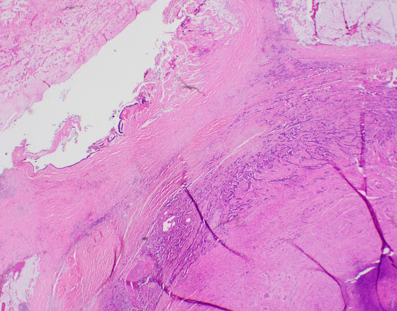
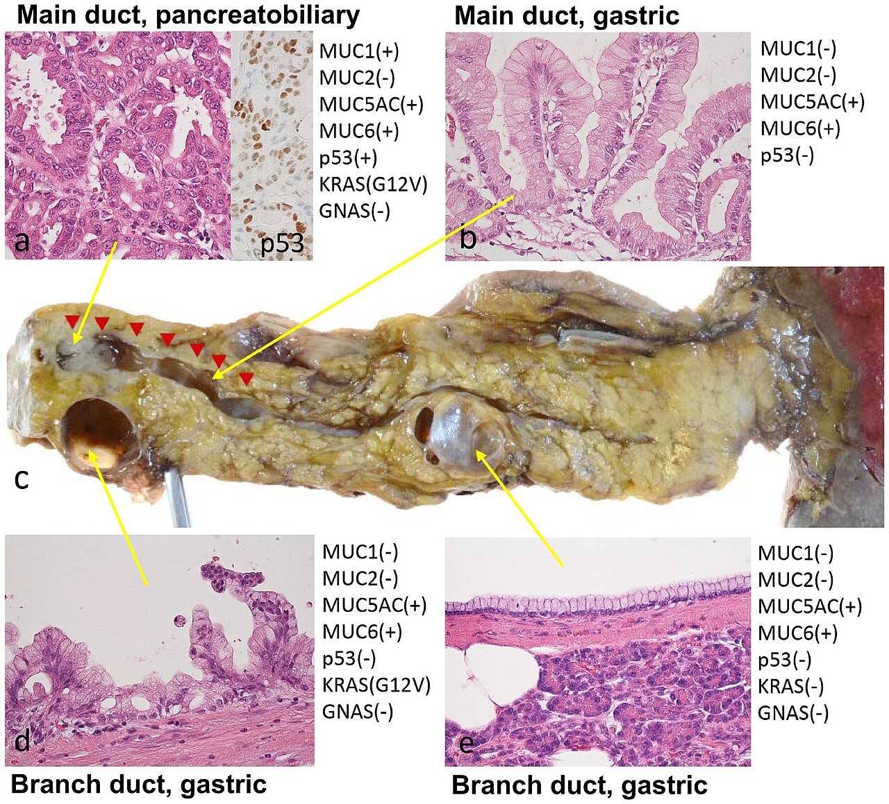
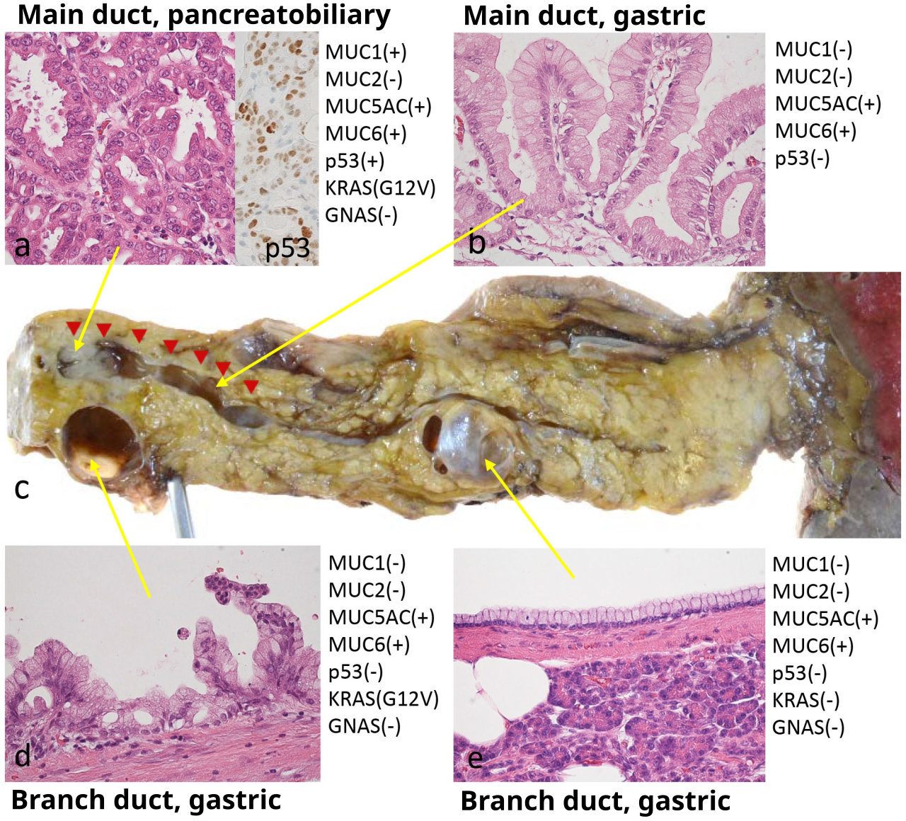
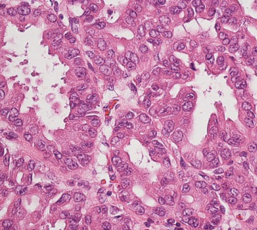
Shibata, Hideki; Ohike, Nobuyuki; Norose, Tomoko; Isobe, Tomohide; Suzuki, Reika · CC BY 4.0
A neoplasm is an abnormal mass of tissue that grows excessively and without control. The process by which this happens is called neoplasia. Unlike normal tissue, a neoplasm’s growth does not coordinate with the surrounding healthy tissue, and it continues to grow even after the initial cause is removed. This growth often results in a lump, commonly referred to as a tumor. The ICD-10 system groups neoplasms into four main categories: benign, in situ, malignant, and those of uncertain or unknown behavior. Malignant neoplasms are also called cancers and are the primary subject of oncology.
Before neoplasia occurs, cells may first show abnormal growth patterns like metaplasia or dysplasia. However, these patterns do not always lead to neoplasia and can appear in other conditions. The word “neoplasm” comes from Ancient Greek, combining *neo* (new) and *plasma* (formation or creation).
Neoplasms can be benign, potentially malignant, or malignant. Benign tumors, such as uterine fibroids, osteophytes, and skin moles, are localized and do not turn into cancer. Potentially malignant neoplasms, like carcinoma in situ, are also localized and do not invade or destroy tissue, but they may become cancerous over time. Malignant neoplasms, or cancers, invade and destroy surrounding tissue, can spread to other parts of the body (metastasize), and are often fatal if not treated. A secondary neoplasm is a cancerous tumor that either spreads from a primary tumor or arises as a new tumor after treatments like chemotherapy or radiotherapy. Rarely, a metastatic neoplasm has no known primary site, a condition called cancer of unknown primary origin.
Neoplastic tumors are often made up of multiple cell types, but their growth typically depends on a single population of neoplastic cells. These cells are thought to be monoclonal, meaning they come from one original cell and share the same genetic or epigenetic abnormality. For lymphoid neoplasms like lymphoma and leukemia, clonality is shown by a single rearrangement of the immunoglobulin gene (in B cell lesions) or T cell receptor gene (in T cell lesions). Proving clonality is now considered essential for identifying a lymphoid cell growth as neoplastic.
The word “tumor” comes from the Latin term for swelling, one of the classic signs of inflammation. Originally, it referred to any swelling, whether neoplastic or not. In modern English, “tumor” is used as a synonym for a neoplasm that forms a solid or fluid-filled mass. However, some neoplasms, like leukemia and most carcinoma in situ, do not form a tumor. Tumor is also not the same as cancer: cancer is always malignant, while a tumor can be benign, precancerous, or malignant. The terms “mass” and “nodule” are often used interchangeably with tumor, but “mass” typically refers to a lesion at least 20 millimeters in its largest dimension, while “nodule” is used for lesions smaller than 20 millimeters.
Tumors in humans result from accumulated genetic and epigenetic changes in single cells, causing them to divide uncontrollably. Not all neoplasms form a tumorous overgrowth—examples include leukemia and carcinoma in situ—but similarities between neoplastic growth and regenerative processes, such as dedifferentiation and rapid cell division, have been noted. Tumor growth has been studied using mathematics and continuum mechanics. Vascular tumors like hemangiomas and lymphangiomas are seen as mixtures of a solid cell skeleton and a liquid filling, allowing researchers to model mechanical stresses and their effects on growth. Experiments show that active tumor growth is often limited to the outer edges, and stiffening of surrounding normal tissue can slow tumor growth.
Benign conditions not involving abnormal tissue growth, such as sebaceous cysts, can also appear as tumors but have no malignant potential. Examples include breast cysts (common during pregnancy) and encapsulated glandular swellings in the thyroid, adrenal gland, or pancreas. Encapsulated hematomas, necrotic tissue from bites or foreign bodies, keloids (overgrowths of scar tissue), and granulomas may also present as tumors. Discrete enlargements of normal structures—like ureters, blood vessels, or bile ducts—due to obstructions, narrowings, or abnormal connections can also mimic tumors. Examples include arteriovenous fistulae, aneurysms, and biliary fistulae.
- field
- Medicine, Oncology
- known_for
- Abnormal tissue growth; classification into benign, potentially malignant, and malignant (cancer); clonality in lymphoid neoplasms
Lore & Background
The word neoplasm is from Ancient Greek νέος- neo 'new' and πλάσμα plasma 'formation, creation'. Prior to the abnormal growth of tissue, cells often undergo metaplasia or dysplasia, though these do not always progress to neoplasia. Neoplasms can be benign, potentially malignant, or malignant. Benign tumors include uterine fibroids, osteophytes, and melanocytic nevi; they are circumscribed and do not transform into cancer. Potentially-malignant neoplasms include carcinoma in situ, which may transform into cancer over time. Malignant neoplasms, commonly called cancer, invade and destroy surrounding tissue, may form metastases, and are generally fatal if untreated.
Reader's Guide
Neoplasms are central to oncology, as malignant neoplasms (cancers) are a leading focus of medical research and treatment. The distinction between benign and malignant tumors guides clinical decisions: benign tumors are localized and non-life-threatening, while malignant tumors require aggressive intervention. The concept of clonality is critical for diagnosing lymphoid neoplasms, where demonstration of a single rearrangement of immunoglobulin or T cell receptor genes confirms neoplastic proliferation. DNA damage is considered the primary underlying cause of malignant neoplasms, with both hereditary and sporadic cancers linked to DNA repair deficiencies. The study of tumor growth using mathematics and continuum mechanics has revealed that active growth is often restricted to the outer edges and that stiffening of normal tissue can inhibit growth. Understanding neoplasms also involves recognizing that not all tumors are neoplastic—benign conditions like cysts, hematomas, and granulomas can present as masses without malignant potential.
Did You Know?
- The word neoplasm comes from Ancient Greek νέος- neo 'new' and πλάσμα plasma 'formation, creation'.
- Some neoplasms, such as leukemia and most forms of carcinoma in situ, do not form a tumor.
- A tumor is not synonymous with cancer; a tumor can be benign, precancerous, or malignant.
- DNA damage is considered the primary underlying cause of malignant neoplasms known as cancers.
Defining the Spectrum of Myeloproliferative Neoplasms
Myeloproliferative neoplasms represent a family of rare blood cancers in which the bone marrow overproduces red blood cells, white blood cells, or platelets beyond the body's needs. The name itself unpacks the pathology: "myelo" points to the marrow, "proliferative" captures the accelerated cell growth, and "neoplasm" signals that this growth is abnormal and uncontrolled. The World Health Organization and the International Consensus Classification jointly maintain the official taxonomy, and as of 2022 they recognize ten distinct entities, ranging from chronic myeloid leukemia and chronic neutrophilic leukemia to polycythemia vera, primary myelofibrosis (split into prefibrotic and overt fibrotic stages), essential thrombocythemia, juvenile myelomonocytic leukemia, chronic eosinophilic leukemia, and a residual category called MPN-NOS. A practical dividing line in clinical practice separates the BCR::ABL1-positive group, which essentially means CML, from all other MPNs. The three conditions most often grouped together as "classical MPNs" are essential thrombocythemia, polycythemia vera, and primary myelofibrosis.
Genetic Origins and the Decades-Long Latency Puzzle
At the cellular level, MPNs begin when precursor blast cells of the myeloid lineages acquire somatic mutations that drive uncontrolled proliferation. The most frequently implicated gene markers include JAK2, CALR, TET2, and MPL, and the specific mutation profile helps clinicians sort patients into the correct subtype. One of the most striking findings in MPN research is that at least some patients already harbor the mutated cell clone in utero, yet the median age at which disease manifests hovers around sixty-two years depending on the subtype. Why a single rogue clone can sit undetectable for decades remains an open question in hematology. Beyond genetics, MPNs share conceptual commonalities with chronic inflammatory diseases, and emerging evidence suggests that environmental exposures may act as additional triggers, much as they do in other long-standing inflammatory conditions. This layered etiology—genetic predisposition meeting possible environmental catalysts—helps explain why the disease landscape is so heterogeneous across patients.
Diagnosing Each Subtype: Markers, Thresholds, and Exclusions
A person with an MPN may be entirely asymptomatic when the condition is first caught on a routine blood panel, which is why the diagnostic workup varies considerably by suspected subtype. Depending on the clinical picture, physicians may order red cell mass determination, a bone marrow aspirate and trephine biopsy, arterial oxygen saturation and carboxyhaemoglobin measurements, neutrophil alkaline phosphatase levels, vitamin B12 or B12-binding capacity assays, serum urate tests, or direct DNA sequencing. Chronic myeloid leukemia is defined by the Philadelphia Chromosome, the BCR::ABL1 fusion. Chronic neutrophilic leukemia requires a CSF3R mutation plus exclusion of other neutrophilia causes. Essential thrombocythemia demands a platelet count above 450 × 10⁹/L, with JAK2 V617F present in up to fifty-five percent of cases and MPL mutations in up to five. Polycythemia vera is tied to JAK2 V617F in over ninety-five percent of patients, with the remainder carrying JAK2 exon 12 mutations, alongside elevated hemoglobin or hematocrit and characteristic marrow findings. The two myelofibrosis stages are distinguished by the degree of reticulin or collagen fibrosis seen on biopsy.
Treatment Realities, Prognosis Shifts, and Rising Incidence
No pharmaceutical cure currently exists for myeloproliferative neoplasms, and for most patients management centers on controlling symptoms and suppressing excessive blood-cell production with myelosuppressive agents. Hematopoietic stem cell transplantation offers a potential cure, but only for a small subset of patients. For essential thrombocythemia and polycythemia vera, the therapeutic priority is preventing thrombohemorrhagic complications, and low-dose aspirin has proven effective in both. In primary myelofibrosis, the goals shift toward alleviating anemia, splenomegaly, and other systemic symptoms. A landmark advance came with tyrosine kinase inhibitors such as imatinib, which lifted CML survival to near-normal life expectancy. More recently, the JAK2 inhibitor ruxolitinib gained approval for primary myelofibrosis, and trials of similar agents are underway for other MPN subtypes. Meanwhile, reported incidence rates are climbing—sometimes tripling—likely reflecting improved genetic diagnostics and refined WHO classification guidelines rather than a true surge in new cases, though wide geographic variation and suspected publication bias for essential thrombocythemia and primary myelofibrosis complicate the picture.
Gallery
Frequently Asked Questions
What is Neoplasm?
Neoplasm refers to an abnormal, excessive proliferation of tissue that grows in a way uncoordinated with the surrounding normal tissue. The process that generates this growth is termed neoplasia, and the resulting mass is commonly called a tumour or tumor.
What are Neoplasm's main classifications?
Neoplasms are sorted into four behavioral categories: benign, in situ, malignant, and those of uncertain or unknown behavior. The malignant subset is what we recognize clinically as cancer.
How does Neoplasm differ from normal tissue growth?
Unlike healthy tissue, a neoplasm keeps proliferating abnormally even after the original triggering stimulus has been eliminated. Its growth pattern is uncoordinated with adjacent normal tissue, which is a hallmark that distinguishes it from physiological regeneration.
What is Neoplasm's role in the medical field?
Neoplasm sits at the heart of oncology, the branch of medicine dedicated to studying and treating abnormal tissue growth. In lymphoid neoplasms specifically, the concept of clonality is a key diagnostic feature used to confirm that the growth originated from a single transformed cell.
Why is Neoplasm important to fans of the Diseases and Conditions series?
As entry 1–15 in the canon, Neoplasm establishes the foundational framework for understanding how abnormal growth ranges from harmless benign masses to life-threatening cancers. Its classification system and behavioral spectrum make it a central reference point for many later entries in the series.
More in Diseases And Conditions 1-15
Spotted an error? Know more?
This is a living reference — every entry is fact-audited, and reader corrections feed straight into our audit queue. Suggest an edit · See this site's audit record