
Urinary system
System that produces and excretes urine in vertebrates.

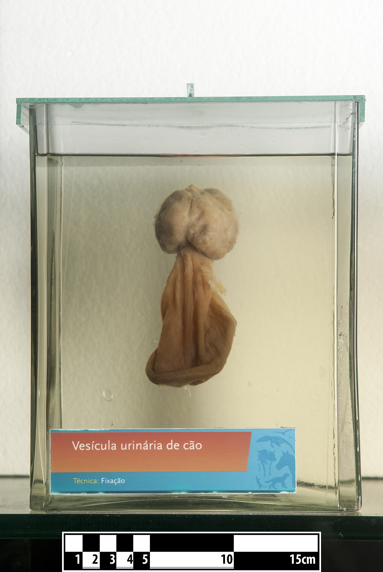
Wagner Souza e Silva · CC BY-SA 4.0
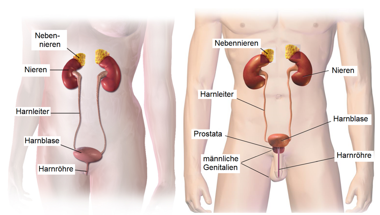
The urinary system, sometimes called the urinary tract or renal system, is part of the vertebrate excretory system. In humans and placental mammals, it includes the kidneys, ureters, bladder, and urethra. Its jobs are to remove urine from the body, manage blood volume and pressure, balance electrolytes and metabolites, and keep blood pH stable. The kidneys get a rich blood supply from the renal arteries, and blood leaves them through the renal vein. Each kidney is made of tiny functional units called nephrons. After blood is filtered and processed, the ureters carry urine to the bladder. The male and female urinary systems are quite alike, with the main difference being urethra length: the male urethra runs through the penis, while the female urethra is shorter and opens near the vulva. A healthy adult typically produces 800 to 2,000 milliliters of urine daily, though this changes with fluid intake and kidney function.
**Structure**
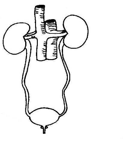
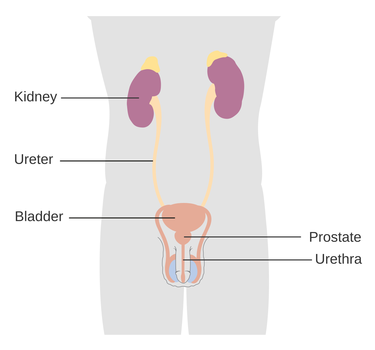
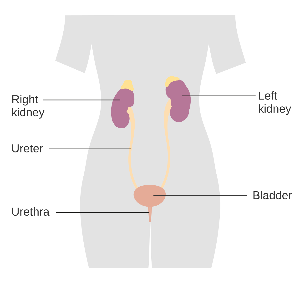
The system includes the organs that make and expel urine. Humans have two kidneys, located on the left and right sides, between the back body wall and the parietal peritoneum. Urine production starts in the nephrons, the kidneys' functional units. From there, urine flows through a series of converging tubules called collecting ducts, which merge into minor calyces, then major calyces, and finally the renal pelvis. The ureters then carry urine to the bladder. The bladder and urethra differ between sexes. In males, the urethra starts at the internal urethral orifice in the bladder's trigone, then passes through the prostatic, membranous, bulbar, and penile sections, ending at the urinary meatus on the glans penis. The female urethra is much shorter, beginning at the bladder neck and opening into the vulval vestibule.
**Microanatomy**
Under a microscope, most of the urinary system—including the renal pelvis, ureters, and bladder—is lined with a special tissue called urothelium, a type of transitional epithelium. Unlike the lining of most organs, this epithelium can flatten and stretch.
**Function**
The urinary system and its parts work to: - Control blood volume and composition (including sodium, potassium, and calcium levels). - Regulate blood pressure. - Maintain blood pH balance. - Help the kidneys produce red blood cells. - Assist in making calcitriol, the active form of vitamin D. - Store waste products (mainly urea and uric acid) before they leave the body.
**Urine formation**
Adults typically produce 1 to 2 liters of urine each day, depending on hydration, activity, environment, weight, and health. Producing too much or too little urine signals a need for medical attention. Polyuria means excessive output (over 2.5 L/day). Low output includes oliguria (under 400 mL/day) and anuria (under 100 mL/day). Urine formation begins with blood filtration in the kidneys. In a healthy person, the kidneys receive 12 to 30% of cardiac output—about 20% on average, or roughly 1.25 L/min. The nephron is the kidney's basic structural and functional unit. Its main role is to balance water and dissolved substances like sodium by filtering blood, reabsorbing what the body needs, and excreting the rest as urine. In the first part of the nephron, Bowman's capsule filters blood from the circulatory system into the tubules. Hydrostatic and osmotic pressure gradients push fluid across a semipermeable membrane. The filtrate contains water, small molecules, and ions that pass easily, while larger molecules like proteins and blood cells are blocked. The amount of filtrate made each minute is the glomerular filtration rate (GFR), totaling about 180 liters per day. Roughly 99% of this filtrate is reabsorbed as it travels through the nephron, and the remaining 1% becomes urine. The endocrine system regulates the urinary system through hormones like antidiuretic hormone, aldosterone, and parathyroid hormone.
**Regulation of concentration and volume**
The circulatory, nervous, and endocrine systems all influence the urinary system. Aldosterone is key for blood pressure control, acting on the distal tubules and collecting ducts of the nephron to increase sodium reabsorption from the filtrate. This sodium reabsorption leads to water retention, raising blood pressure and volume. Antidiuretic hormone (ADH), also called vasopressin, is a neurohypophysial hormone found in most mammals. Its two main jobs are to retain water in the body and cause vasoconstriction. Vasopressin boosts water reabsorption in the kidney's collecting ducts by moving aquaporin-CD water channels into the plasma membrane of those duct cells.
**Urination**
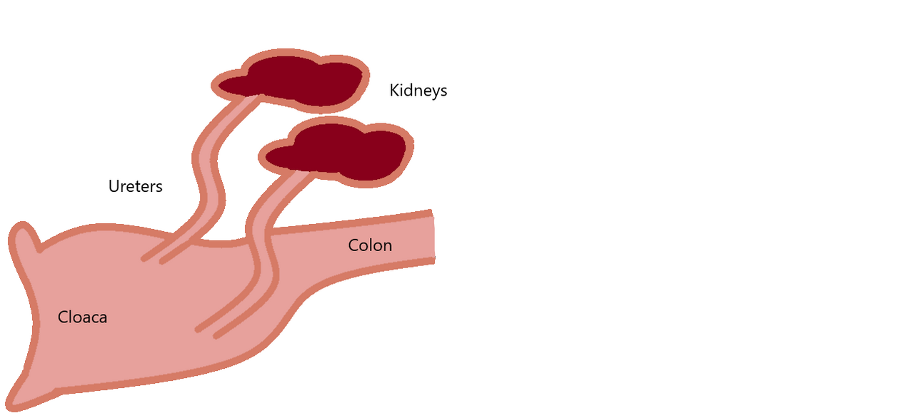
Urination, also known as micturition, is the release of urine from the bladder to the outside. In placental mammals, urine exits through the urethra from the penis or vulva; in other vertebrates, it passes through the cloaca. In healthy humans (and many other animals), urination is under voluntary control. In infants, some elderly people, and those with nerve damage, urination may happen involuntarily.
- field
- Anatomy and physiology
- known_for
- Elimination of urine, regulation of blood volume, blood pressure, electrolyte balance, and blood pH
- components
- Kidneys, ureters, bladder, urethra
Lore & Background
The urinary system refers to the structures that produce and excrete urine. In the human urinary system there are two kidneys located between the dorsal body wall and parietal peritoneum on both the left and right sides. The formation of urine begins within the functional unit of the kidney, the nephrons. Urine then flows through the nephrons, through a system of converging tubules called collecting ducts, which join to form the minor calyces, followed by the major calyces that ultimately join the renal pelvis, followed by the ureters, which transport urine into the urinary bladder. The anatomy of the human urinary bladder and urethra differs between males and females. In males, the urethra begins at the internal urethral orifice in the trigone of the bladder, and then becomes the prostatic, membranous, bulbar, and penile urethra, terminating at the urinary meatus in the glans penis. The female urethra is much shorter, beginning at the bladder neck and terminating in the vulval vestibule.
Reader's Guide
The urinary system is significant for its role in maintaining homeostasis through the regulation of blood volume, blood pressure, electrolyte levels, and blood pH. It also contributes to red blood cell production and the synthesis of calcitriol, the active form of Vitamin D. The system stores waste products such as urea and uric acid before elimination. Urine formation begins with filtration of blood in the kidneys, where the nephrons regulate water and soluble substances. The system is regulated by the endocrine system via hormones like antidiuretic hormone, aldosterone, and parathyroid hormone. Clinical significance includes urologic diseases such as urinary tract obstruction, kidney tissue diseases treated by nephrologists, and urinary tract diseases treated by urologists. Urinary incontinence can result from weakening of the pelvic floor muscles, and behavioral therapy generally improves outcomes. Cancers of the urinary system include bladder, kidney, ureteral, and urethral cancer. Historically, kidney stones have been recorded since ancient times, and the ureters' function was described by Galen in the second century AD.
Did You Know?
- The kidneys receive about 20% of cardiac output, averaging about 1.25 L/min.
- The urinary system is covered in a unique lining called urothelium, a type of transitional epithelium that can flatten and distend.
Architecture of the Urinary Tract
The urinary system in humans and placental mammals is built around four principal organs: a pair of kidneys, two ureters, a bladder, and a urethra. The kidneys sit against the dorsal body wall, nestled between the parietal peritoneum on either side of the spine. Each kidney is packed with millions of nephrons, the tiny functional units where urine is actually manufactured. Once formed, urine travels through a cascade of converging tubules known as collecting ducts, which feed into minor calyces, then major calyces, and finally the renal pelvis. From there, the ureters carry the fluid down into the bladder for temporary storage. The final exit route, the urethra, is where male and female anatomy diverge most noticeably. In males, the urethra is a long, multi-segmented channel passing through the prostatic, membranous, bulbar, and penile regions before opening at the glans. In females, the urethra is considerably shorter, running from the bladder neck to the vulval vestibule. This structural difference in urethral length is the primary anatomical distinction between the sexes' urinary tracts.
The Filtration Engine: How Urine Is Made
Urine production begins with a remarkable act of selective filtering. In a healthy adult, the kidneys receive roughly twenty percent of the heart's total output, about 1.25 litres of blood every minute. Within each nephron, Bowman's capsule acts as the first checkpoint, using hydrostatic and osmotic pressure gradients to push water, small molecules, and ions across a semipermeable membrane into the tubule lumen. Larger entities such as proteins and blood cells are too big to cross and remain in the bloodstream. This initial filtrate amounts to a staggering 180 litres per day, yet the nephron reabsorbs approximately ninety-nine percent of it as it passes through successive segments. Only that remaining one percent exits as urine, typically 800 to 2,000 millilitres per day in a healthy person. The entire process is fine-tuned by the endocrine system, with hormones like antidiuretic hormone, aldosterone, and parathyroid hormone modulating how much water and which electrolytes are reclaimed versus excreted.
Homeostatic Guardian: Beyond Waste Removal
While eliminating urea and uric acid is the most visible role of the urinary system, its deeper significance lies in maintaining internal balance. The kidneys actively regulate blood volume and the concentrations of critical electrolytes such as sodium, potassium, and calcium. They also serve as a barostat for blood pressure: aldosterone, acting on the distal tubules and collecting ducts, prompts reabsorption of sodium, which in turn drags water back into circulation, raising both volume and pressure. Antidiuretic hormone, or vasopressin, reinforces water retention by triggering the insertion of aquaporin-CD channels into the collecting duct membrane, making the tubule more permeable to water. Beyond fluid balance, the kidneys contribute to red blood cell production and help synthesize calcitriol, the biologically active form of vitamin D. They also buffer blood pH, keeping the internal environment within a narrow, survivable range. In this way, the urinary system functions as a multifaceted regulatory organ rather than a simple waste-disposal pipeline.
From Bladder to Outside: Urination and Clinical Watchpoints
The act of emptying the bladder, known as urination or micturition, is a coordinated neurological event. In healthy adults it is a voluntary process, but in infants, some elderly individuals, and people with neurological injury it can occur as an involuntary reflex. The brain orchestrates the sequence through the pontine micturition center, the periaqueductal gray, and the cerebral cortex, integrating central, autonomic, and somatic signals to relax the bladder wall while contracting the urethral sphincters. The lining that makes this system work is the urothelium, a specialized transitional epithelium covering the renal pelvis, ureters, and bladder. Unlike the flat epithelia of most organs, urothelium can stretch and flatten as the bladder fills, then compress when it empties. Clinically, urine output is a vital diagnostic window: more than 2.5 litres a day signals polyuria, less than 400 millilitres indicates oliguria, and under 100 millilitres points to anuria, each demanding medical investigation.
Gallery
Frequently Asked Questions
Who is Urinary system?
The urinary system is a vertebrate organ network dedicated to waste removal and fluid balance. In humans and placental mammals it is assembled from four main structures: the kidneys, ureters, bladder, and urethra.
What are Urinary system's powers/role?
It filters blood to produce urine, keeps blood volume and pressure within safe ranges, and fine-tunes electrolyte and metabolite concentrations. It also serves as the body's primary pH regulator, holding the internal environment in a stable state.
How does Urinary system's story end?
Its narrative arc concludes with the excretion of urine out of the body through the urethra. That final step closes the full cycle of filtration, reabsorption, and waste elimination.
Why is Urinary system important?
Without it, vertebrates would lose the ability to maintain stable blood pressure, fluid volume, and ionic balance. It is the principal mechanism for keeping blood pH within the narrow range that living cells require to survive.
What team or guild does Urinary system belong to?
It operates as a subsystem within the broader excretory system of vertebrates. Its field of operation spans anatomy and physiology, with a specific focus on renal function and urine production.
More in Physiology & Medicine 1-19
Spotted an error? Know more?
This is a living reference — every entry is fact-audited, and reader corrections feed straight into our audit queue. Suggest an edit · See this site's audit record