
Stomach
A muscular digestive organ between esophagus and small intestine.

Calicut Medical College · CC BY-SA 4.0
The stomach is a muscular, hollow organ found in the upper gastrointestinal tract of humans, many other animals, and even some invertebrates. Its ancient Greek name, *gaster*, gives us the medical term "gastric." This dilated structure is a key part of the digestive system, playing a central role in the gastric phase of digestion—the stage that follows the cephalic phase, which is triggered by seeing, smelling, or chewing food. Inside the stomach, food is chemically broken down by digestive enzymes and gastric acid. The organ also helps regulate gut microbiota, which influences both digestion and overall health.
Situated between the esophagus and the small intestine, the stomach uses the pyloric sphincter to control the release of partially digested food, called chyme, into the duodenum—the first and shortest section of the small intestine. From there, peristalsis moves the chyme through the rest of the intestines.
**Structure** In humans, the stomach lies between the esophagus and the duodenum, positioned in the left upper quadrant of the abdominal cavity. Its top rests against the diaphragm, while the pancreas sits behind it. A large double fold of visceral peritoneum, the greater omentum, hangs down from the stomach’s greater curvature. Two sphincters keep stomach contents contained: the lower esophageal sphincter (in the cardiac region) at the junction with the esophagus, and the pyloric sphincter at the junction with the duodenum. The stomach is surrounded by parasympathetic (inhibitory) and sympathetic (stimulatory) nerve networks—plexuses in the anterior gastric, posterior, superior, inferior, celiac, and myenteric regions—that regulate both its secretion and muscle movement. The organ is distensible, normally expanding to hold about one liter of food. Its shape depends on how full it is and the state of nearby organs, like the colon. When empty, it is somewhat J-shaped; when partially distended, it becomes pear-shaped. In obese individuals, it tends to be more horizontal. A newborn’s stomach holds only about 30 milliliters, while adult maximum volume ranges from 2 to 4 liters, though volumes up to 15 liters have been recorded in extreme cases.
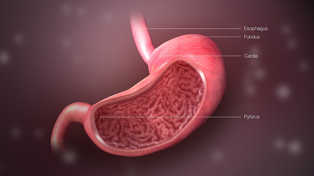
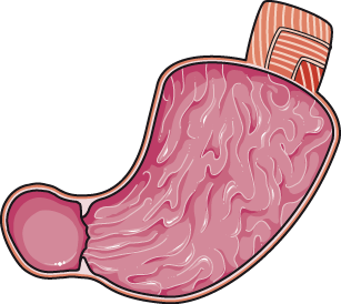
**Sections** The human stomach has four sections: cardia, fundus, body, and pylorus. The cardia is a small region surrounding the esophageal opening, where contents from the esophagus enter through the cardiac orifice. A cardiac notch on the left marks the start of the greater curvature. A horizontal line from this notch defines the dome-shaped fundus (from Latin for "bottom"). The body, or corpus, is the main central region. The pylorus (from Greek for "gatekeeper") connects the stomach to the duodenum via the pyloric sphincter, with the pyloric antrum opening into the body. The cardia follows the "z-line" of the gastroesophageal junction, where the epithelium changes from stratified squamous to columnar; the lower esophageal sphincter lies nearby.
**Anatomical Proximity** The "stomach bed" refers to the structures the stomach rests upon in mammals, including the tail of the pancreas, splenic artery, left kidney, left suprarenal gland, transverse colon and its mesocolon, left crus of the diaphragm, and left colic flexure. This term was introduced around 1896 by Philip Polson of the Catholic University School of Medicine, Dublin, though it was later discredited by surgeon anatomist J. Massey.
**Blood Supply** The lesser curvature receives blood from the right gastric artery (inferiorly) and left gastric artery (superiorly), with the left also supplying the cardiac region. The greater curvature is supplied by the right gastroepiploic artery (inferiorly) and left gastroepiploic artery (superiorly). The fundus and upper greater curvature are supplied by 5 to 7 short gastric arteries arising from the splenic artery.
**Lymphatic Drainage** Two sets of gastric lymph nodes drain the stomach’s tissue fluid into the lymphatic system via the intestinal lymph trunk, reaching the cisterna chyli.
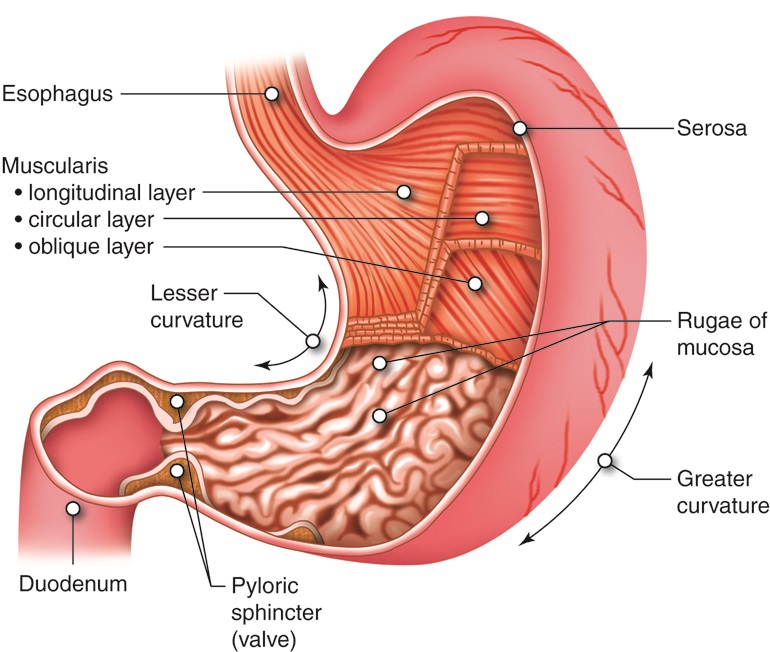
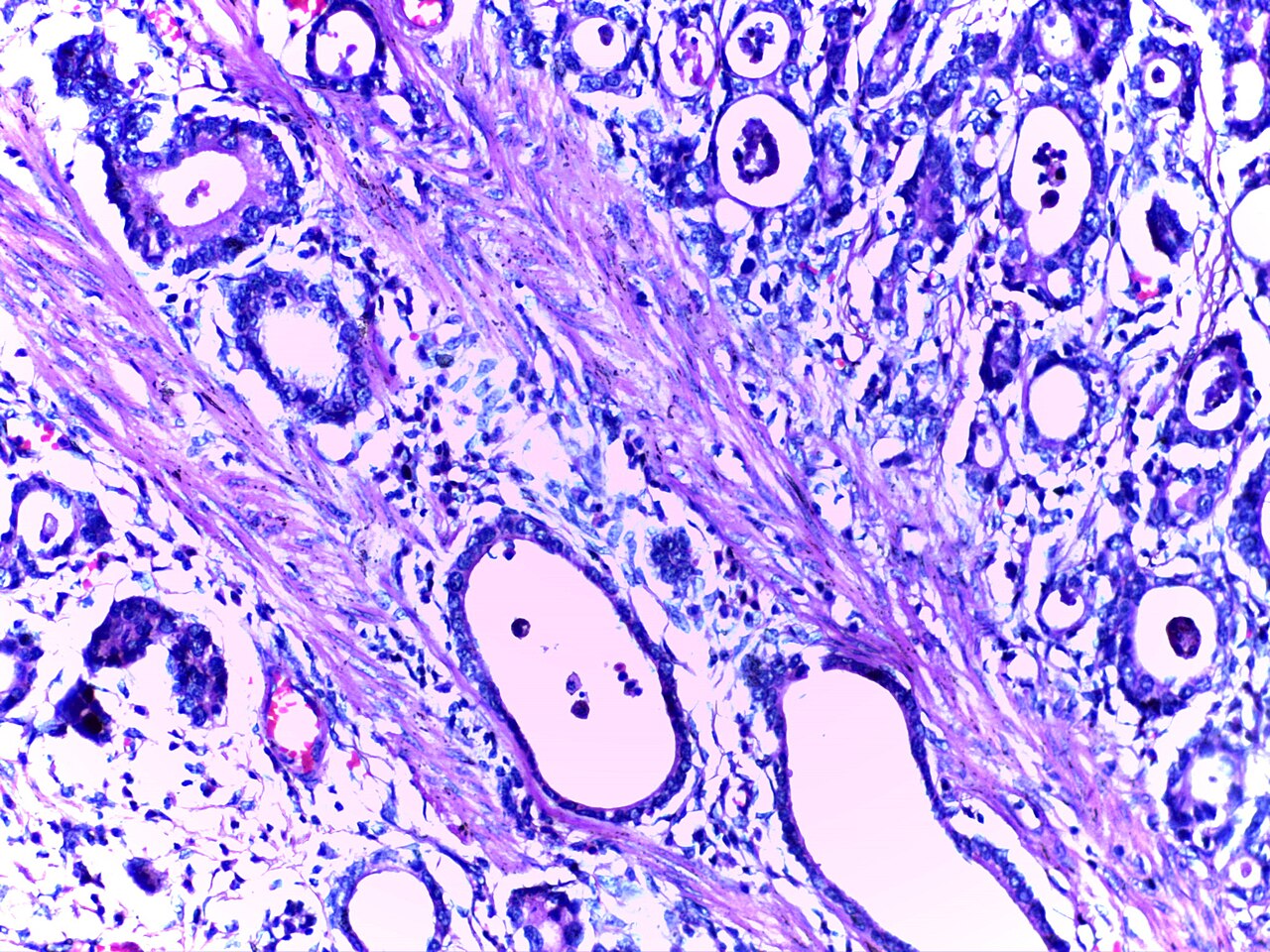
**Microanatomy** Like other parts of the gastrointestinal wall, the human stomach wall consists, from inner to outer, of a mucosa, submucosa, muscular layer, subserosa, and serosa. The inner gastric mucosa is a mucous membrane lining the stomach, made of an outer columnar epithelium, a lamina propria, and a thin smooth muscle layer called the muscularis mucosa. Beneath this lies the submucosa, composed of fibrous connective tissue, containing Meissner’s plexus interior to the oblique muscle layer. Outside the submucosa is the muscular layer, which has three layers of muscle fibers lying at angles to each other: inner oblique, middle circular, and outer longitudinal. The inner oblique layer is unique to the stomach, not found elsewhere in the gastrointestinal tract. The stomach has the thickest muscular layer of the tract, with three layers, allowing maximum peristalsis. The inner oblique layer creates the churning motion that physically breaks down food.
- Capacity
- Normally about one litre; maximum adult volume 2–4 litres; up to 4–6 litres in extreme pathological distension
Lore & Background
The stomach lies between the esophagus and the duodenum, the first part of the small intestine. It is located in the left upper quadrant of the abdominal cavity, with its top against the diaphragm and the pancreas behind it. A large double fold of visceral peritoneum called the greater omentum hangs down from the greater curvature. Two sphincters—the lower esophageal sphincter at the cardiac region and the pyloric sphincter at the duodenal junction—keep stomach contents contained. The stomach is surrounded by parasympathetic and sympathetic plexuses that regulate secretory and motor activity.
Reader's Guide
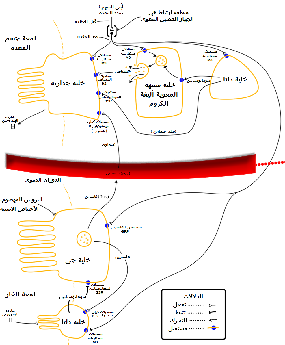
The stomach is distensible, normally expanding to hold about one litre of food, with a shape that depends on distension and surrounding viscera; when empty it is somewhat J-shaped, and when partially distended it becomes pyriform. In obese persons it is more horizontal, and a newborn's stomach holds only about 30 millilitres. The human stomach can be divided into four sections: cardia, fundus, body, and pylorus. The gastric cardia receives contents from the esophagus; the fundus is the dome-shaped upper part; the body is the main central region; and the pylorus connects to the duodenum via the pyloric sphincter. The stomach wall consists of mucosa, submucosa, muscular layer, subserosa, and serosa, with the muscular layer having three layers—inner oblique, middle circular, and outer longitudinal—the inner oblique being unique to the stomach. Gastric glands secrete hydrochloric acid, digestive enzymes, mucus, and hormones such as gastrin.
Did You Know?
- The Ancient Greek name for the stomach is gaster, used in medical terms such as gastric.
- The stomach can normally expand to hold about one litre of food, but volumes up to 4–6 litres have been observed in extreme pathological distension.
- The inner oblique muscle layer of the stomach is not seen in other parts of the digestive system.
- The pylorus is named from the Greek word for 'gatekeeper'.
Anatomical Architecture & Positioning
The stomach occupies the left upper quadrant of the abdominal cavity, nestled between the esophagus above and the duodenum below. Its dome-shaped fundus presses against the diaphragm, while the pancreas lies directly behind it. A large double fold of visceral peritoneum, the greater omentum, drapes from the greater curvature. The organ is divided into four sequential regions: the tiny cardia surrounding the esophageal opening, the fundus forming the upper curved dome, the body or corpus as the main central chamber, and the pylorus connecting to the duodenum. The cardia is marked by the z-line where epithelium transitions from stratified squamous to columnar. The stomach bed—the structures upon which the organ rests—includes the pancreatic tail, splenic artery, left kidney, left suprarenal gland, transverse colon with its mesocolon, the left crus of the diaphragm, and the left colic flexure. This term was coined around 1896 by Philip Polson at the Catholic University School of Medicine in Dublin, though it was later challenged by the surgeon-anatomist J Massey.
The Muscular Engine & Peristalsis
What sets the stomach apart from the rest of the gastrointestinal tract is its uniquely thick muscular wall, composed of three distinct layers of smooth muscle fibers arranged at angles to one another. The inner oblique layer is found nowhere else in the digestive system and is responsible for the powerful churning motion that physically breaks down food. The middle circular layer forms a thick, tonically constricted ring at the pylorus, creating a functional sphincter that regulates the release of chyme. The outer longitudinal layer completes the triad. Because of this triple-layered architecture, the stomach generates the maximum peristaltic force in the entire gut. Two sphincters govern its contents: the lower esophageal sphincter at the cardiac junction prevents backflow into the esophagus, while the pyloric sphincter at the duodenal junction meters out partially digested material. The antrum's walls contain thicker muscle cells and contract more forcefully than the fundus, giving the organ a gradient of mechanical power from top to bottom.
Digestive Function & Chemical Processing
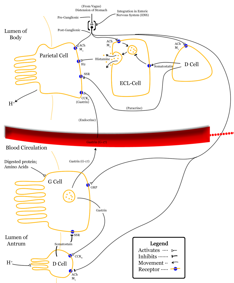
The stomach serves as the central stage of the gastric phase of digestion, which follows the cephalic phase triggered by the sight, smell, and chewing of food. Within its lumen, secreted digestive enzymes and gastric acid work together to chemically break down ingested material. The organ also plays a role in regulating gut microbiota, thereby influencing broader digestive processes and overall health. Once food has been sufficiently processed, the pyloric sphincter releases the resulting chyme into the duodenum—the first and shortest segment of the small intestine—where peristalsis takes over to propel it through the remaining intestinal tract. The stomach's capacity is remarkably variable: a newborn's stomach holds roughly 30 millilitres, a typical adult can accommodate about one litre, and maximum adult volume ranges from two to four litres, with extreme observations reaching as high as fifteen litres. Its shape shifts with distension—J-shaped when empty, pyriform when partially filled, and more horizontal in obese individuals.
Vascular & Neural Infrastructure
The stomach's blood supply is distributed across its curvatures by a network of arteries. The lesser curvature receives the right gastric artery inferiorly and the left gastric artery superiorly, the latter also feeding the cardiac region. The greater curvature is served by the right gastroepiploic artery below and the left gastroepiploic artery above, while the fundus and upper greater curvature are nourished by five to seven short gastric arteries branching from the splenic artery. Lymphatic drainage flows through two sets of gastric lymph nodes into the intestinal lymph trunk and ultimately the cisterna chyli. Neural regulation is equally elaborate: parasympathetic (inhibitory) and sympathetic (stimulatory) plexuses—including the anterior gastric, posterior, superior, inferior, celiac, and myenteric networks—wrap around the organ to coordinate both its secretory and motor activities. This dual innervation ensures that the stomach's chemical and mechanical work is finely tuned to the body's changing needs.
Gallery
Frequently Asked Questions
Who is Stomach?
The stomach is a J-shaped muscular chamber in the upper abdomen, sitting between the esophagus and the small intestine. It acts as the central mixing and chemical-digestion hub of the gastrointestinal tract.
What are Stomach's powers/role?
It churns ingested food while releasing hydrochloric acid and pepsin to break down proteins during the gastric phase of digestion. It also helps shape the gut microbiome, which in turn influences nutrient absorption and overall systemic health.
How does Stomach's story end?
In a healthy adult the gastric lining renews itself continuously, so the organ does not simply wear out like a blood cell. Chronic insults such as H. pylori infection or long-term NSAID use, however, can erode that lining and progress to ulcers or, in rare cases, gastric carcinoma.
Why is Stomach important?
Without its acidic milieu and enzymatic action, dietary proteins would reach the small intestine largely intact, depriving the body of usable amino acids. It also paces the release of chyme so the downstream intestine is never flooded all at once.
How big is Stomach?
Under everyday conditions it holds roughly one litre of contents, but a healthy adult can comfortably accommodate two to four litres. In extreme pathological distension, volumes of four to six litres have been recorded before the risk of rupture becomes critical.
More in Physiology & Medicine 1-19
Spotted an error? Know more?
This is a living reference — every entry is fact-audited, and reader corrections feed straight into our audit queue. Suggest an edit · See this site's audit record