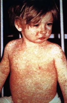
Histiocytosis X
A clonal proliferation of Langerhans cells affecting skin, bone, and other organs.
Last updated

via Wikipedia: Langerhans cell histiocytosis · see source
Histiocytosis X is an older name for Langerhans cell histiocytosis (LCH), a disorder marked by abnormal clonal proliferation of Langerhans cells derived from bone marrow. It is part of the histiocytoses, a group of syndromes involving abnormal proliferation of histiocytes (an archaic term for activated dendritic cells and macrophages), and is related to other white blood cell proliferations such as leukemias and lymphomas. The disease spectrum ranges from isolated bone lesions to multisystem disease. The name histiocytosis X was used until 1985, when the Histiocyte Society renamed it Langerhans cell histiocytosis.
Quick Facts
- Field
- Hematology
Facts from the source article.
Classification
Histiocytosis X, also known as Langerhans cell histiocytosis (LCH), involves the abnormal buildup of cells that look like Langerhans cells, a type of dendritic cell in the skin. These cells, along with lymphocytes, eosinophils, and normal histiocytes, create lesions that can appear in almost any organ. A similar condition has been described in canine histiocytic diseases. LCH is split into three clinical types: unifocal, multifocal unisystem, and multifocal multisystem.
Unifocal
Unifocal LCH, once called eosinophilic granuloma (a term now known to be a misnomer), features a growing mass of Langerhans cells in a single organ, causing damage. It typically has no extraskeletal involvement, but rarely a lesion can be found in the skin, lungs, or stomach. It can appear as a single lesion in an organ, up to a large quantity of lesions in one organ. When multiple lesions are scattered throughout an organ, it can be called a multifocal unisystem variety.
When found in the lungs, it should be distinguished from Pulmonary Langerhans cell histiocytosis—a special category most commonly seen in adult smokers. When found in the skin it is called cutaneous single system Langerhans cell LCH; this version can heal without therapy in some rare cases.
Multifocal unisystem
This primary bone involvement helps to differentiate eosinophilic granuloma from other forms of LCH (Letterer-Siwe or Hand-Schüller-Christian variant). Multifocal unisystem LCH is seen mostly in children, characterized by fever, bone lesions and diffuse eruptions, usually on the scalp and in the ear canals. 50% of cases involve the pituitary stalk, often leading to diabetes insipidus.
Multifocal multisystem
The triad of diabetes insipidus, exophthalmos, and lytic bone lesions is known as the Hand-Schüller-Christian triad. Peak onset is 2–10 years of age. Multifocal multisystem LCH, also called Letterer-Siwe disease, is an often rapidly progressing disease in which Langerhans cells proliferate in many tissues. It is mostly seen in children under age 2, and the prognosis is poor: even with aggressive chemotherapy, the five-year survival is only 50%.
Signs and symptoms
LCH provokes a non-specific inflammatory response, which includes fever, lethargy, and weight loss. The most frequently seen symptom in both unifocal and multifocal disease is painful bone swelling. The skull is most frequently affected, followed by the long bones of the upper extremities and flat bones. Infiltration in hands and feet is unusual.
Osteolytic lesions can lead to pathological fractures. Commonly seen skin findings are a rash which varies from scaly erythematous lesions to red papules pronounced in intertriginous areas. Up to 80% of LCH patients have extensive eruptions on the scalp. Bone marrow involvement can cause pancytopenia with superadded infection, usually implying a poor prognosis.
Anemia can be due to a number of factors and does not necessarily imply bone marrow infiltration. Lymph node enlargement occurs in 50% of cases, spleen enlargement in 30%, and liver enlargement in 20% of Histiocytosis cases.
Endocrine involvement commonly affects the hypothalamic-pituitary axis; central diabetes insipidus can be the first manifestation of LCH, and anterior pituitary hormone deficiency is usually permanent. Lung involvement may be asymptomatic, diagnosed incidentally because of lung nodules on radiographs, or cause chronic cough and shortness of breath. Less frequently, the gastrointestinal tract, central nervous system, and oral cavity are affected.
Pathophysiology
The pathogenesis of LCH is a matter of debate, with ongoing investigations to determine whether it is a neoplastic (cancerous) or reactive (non-cancerous) process. Arguments supporting the reactive nature include the occurrence of spontaneous remissions, the extensive secretion of multiple cytokines by dendritic cells and bystander-cells (a phenomenon known as cytokine storm) in the lesional tissue, favorable prognosis, and relatively good survival rate in patients without organ dysfunction or risk organ involvement.
On the other hand, the infiltration of organs by monoclonal population of pathologic cells, and the successful treatment of subset of disseminated disease using chemotherapeutic regimens are consistent with a neoplastic process. In addition, a demonstration, using X chromosome–linked DNA probes, of LCH as a monoclonal proliferation provided additional support for the neoplastic origin of this disease. While clonality is an important attribute of cancer, its presence does not prove that a proliferative process is neoplastic.
Recurrent cytogenetic or genomic abnormalities would also be required to demonstrate convincingly that LCH is a malignancy. An activating somatic mutation of a proto-oncogene in the Raf family, the BRAF gene, was detected in 35 of 61 (57%) LCH biopsy samples, with mutations being more common in patients younger than 10 years (76%) than in patients aged 10 years and older (44%).
This study documented the first recurrent mutation in LCH samples. Two independent studies have confirmed this finding. Presence of this activating mutation could support the notion to characterize LCH as a myeloproliferative disorder.
Diagnosis
Diagnosis is confirmed histologically by tissue biopsy. Hematoxylin-eosin stain of biopsy slide will show features of Langerhans cells, e.g., distinct cell margin, pink granular cytoplasm. Presence of Birbeck granules on electron microscopy and immunocytochemical features, e.g., CD1 positivity, are more specific. Initially routine blood tests, e.g., full blood count, liver function test, U&Es, bone profile, are done to determine disease extent and rule out other causes.
Electron microscopy or immunohistochemistry (only one of the two is required) may be used to definitively diagnose LCH, in the presence of other pathologic and histologic qualities of the disease. Imaging may be evident in chest X-rays with micronodular and reticular changes of the lungs with cyst formation in advanced cases. MRI and high-resolution CT may show small, cavitated nodules with thin-walled cysts. MRI scan of the brain can show three groups of lesions: tumourous/granulomatous lesions, nontumourous/granulomatous lesions, and atrophy.
Tumourous lesions are usually found in the hypothalamic-pituitary axis with space-occupying lesions with or without calcifications. In non-tumourous lesions, there is a symmetrical hyperintense T2 signal with hypointense or hyperintense T1 signal extending from grey matter into the white matter.
Did You Know?
- A very rare self-limiting variant seen at birth is called Hashimoto-Pritzker disease.
- Pulmonary Langerhans cell histiocytosis occurs almost exclusively in cigarette smokers and is now considered a form of smoking-related interstitial lung disease.
More in Monocyte- and Macrophage-related Cutaneous Conditions
Related in Monocyte- and Macrophage-related Cutaneous Conditions
Links follow this subject's own source article.
Sources
Compiled from Wikipedia and the sources listed below. Text from Wikipedia is available under CC BY-SA 4.0; this entry is adapted from it.
- Wikipedia: Langerhans cell histiocytosis (CC BY-SA 4.0).
- Word definitions: the , each quoted from its Wikipedia article.
Spotted an error? Know more?
Reader corrections go straight into our review queue. Suggest an edit · How this site is sourced