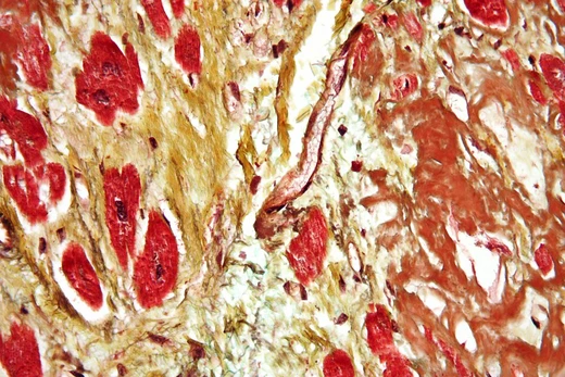
Cardiac amyloidosis
Protein deposits stiffen the heart, causing restrictive failure.
Last updated
Cardiac amyloidosis is a subcategory of amyloidosis in which the misfolded protein amyloid accumulates in the heart muscle, valves, or atria, causing thickening and reduced cardiac function. This multisystem disease was often misdiagnosed and only confirmed at autopsy, but advances in technology have enabled earlier detection. It encompasses several subtypes, including light chain, hereditary transthyretin, and wild-type transthyretin amyloidosis.
Types
The multiple subtypes of cardiac amyloidosis have varying epidemiological, diagnostic, and prognostic characteristics. Seventy-five percent of transthyretin (TTR) cardiac amyloidosis is the wild-type and 25% is the inherited type. Light chain cardiac amyloidosis is a relatively rare form occurring in an estimated six to ten cases per 1,000,000 people; it usually affects males over age 60 and is rapidly progressive. Its pathogenesis involves aggregation of immunoglobulin lambda light chains produced by an abnormal expansion of plasma cells, which deposit into myocardial interstitial tissue.
Diagnostic tests include serum and urine electrophoresis, elevated troponin and BNP levels, and ECGs showing low QRS voltages. Familial (ATTRm) cardiac amyloidosis is caused by mutations in the TTR gene, with approximately 130 known variants; incidence varies by population. A common mutation, Val122Ile, is estimated to be present in 3.5–4% of African Americans in the United States. This type can be identified by genetic testing, and histological evaluation uses stains such as Congo Red, which shows apple green birefringence under polarized light.
Familial amyloidosis usually causes heart failure combined with peripheral or autonomic neuropathy. Wild-type transthyretin amyloidosis has no TTR gene mutations but still forms deposits in the heart and other tissues, affecting males over age 70. Carpal tunnel syndrome, tendon rupture, or spinal stenosis may precede heart failure by 10–15 years.
Diagnosis
Echocardiography is a safe, non-invasive method to assess structural and functional heart disease. In amyloidosis, it reveals ventricle and valvular thickening, biatrial enlargement, restrictive filling, normal to mildly reduced systolic function, and decreased diastolic filling. A specific pattern—relative preservation of apical myocardium with decreased longitudinal strain in mid and basal sections—is 90–95% sensitive and 80–85% specific for cardiac amyloidosis, though echocardiography is not definitive.
ECGs typically show low voltage in limb leads, an unusual extreme right axis, and a normal or slightly prolonged P-wave. In light-chain amyloidosis, the QRS complex has poor R-waves in chest leads; in TTR amyloidosis, there is discordance between wall thickness suggested by EKG and actual wall thickness. Holter monitors can identify asymptomatic arrhythmias. Atrial fibrillation is observed in up to 70% of patients at diagnosis, often with controlled ventricular rates due to conduction system disease.
Laboratory tests include urea, creatinine, liver enzymes, glucose, thyroid function, full blood count, clotting tests, and serum and urine immunofixation to detect monoclonal bands consistent with light chain amyloidosis. Serum immunoglobulin free light chain assay aids diagnosis and monitoring.
Treatments
Treatments differ according to the type of amyloidosis. Most aim to preserve heart function and manage heart failure symptoms, with loop diuretics used for fluid overload. For light chain (AL-CM) amyloidosis, the major goal is reducing free light chain concentration, often by killing the abnormal plasma cells with chemotherapeutic agents such as melphalan or bortezomib. Following chemotherapy, a bone marrow transplant can restore normal cell lines.
Newer medications under research for multiple myeloma (ixazomib, carfilzomib, daratumumab, elotuzumab) may also decrease free light chain production. Orthotopic heart transplant followed by melphalan and stem cell transplant has shown results similar to non-cardiac amyloidosis heart transplant.
Complications may be treated with midodrine for autonomic neuropathy, amiodarone for atrial fibrillation, and warfarin after a cardioembolic episode. For familial (ATTRm-CM) amyloidosis, recent developments include methods to suppress transthyretin production, stabilize amyloid fibrils, and destroy existing fibrils. ACE-inhibitors and beta-blockers can be prescribed if autonomic neuropathy is absent.
Prognosis
Overall prognosis depends on the extent of cardiac dysfunction; worse outcomes are seen when echocardiography shows left ventricular wall thickness, poor systolic function, and severe diastolic dysfunction. For light-chain amyloidosis, early detection offers the best chance of prolonged remission, with well-treated cases having a 4-year survival rate of around 90%. Patients undergoing stem cell transplant have an average survival time of 10 years.
Staging systems such as the Mayo Biomarker Stage use troponin I, troponin T, BNP, NT-proBNP, and free light chain concentrations. For familial (ATTRm-CM) amyloidosis, prognosis varies by mutation type and age of onset; it is generally slower to progress than light chain amyloidosis. However, the Val122Ile mutation has a 4-year survival rate of 16% with an average length of 26 months, and a delay in recognition contributes to this reduced survival.
Did You Know?
- Macroglossia and periorbital bruising are extracardiac manifestations of light-chain amyloidosis.
- The P component of amyloid stabilizes fibrils, reducing their clearance from the body.
Frequently Asked Questions
What are the symptoms of Cardiac amyloidosis?
Symptoms of Cardiac amyloidosis include Shortness of breath, fatigue, swelling in legs and ankles and irregular heartbeat.
How is Cardiac amyloidosis diagnosed?
Diagnosis of Cardiac amyloidosis is based on echocardiogram, cardiac MRI, biopsy and nuclear imaging.
More in Heart Diseases
Sources
Compiled from Wikipedia and the sources listed below. Text from Wikipedia is available under CC BY-SA 4.0; this entry is adapted from it.
- Wikipedia: Cardiac amyloidosis (CC BY-SA 4.0).
- Word definitions: the , each quoted from its Wikipedia article.
Spotted an error? Know more?
Reader corrections go straight into our review queue. Suggest an edit · How this site is sourced