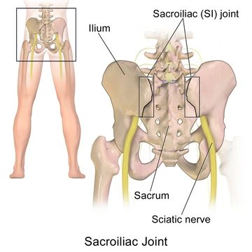
Sacroiliac joint dysfunction
Abnormal sacroiliac joint motion causing pain.
Sacroiliac joint dysfunction is a condition characterized by abnormal motion in the sacroiliac joint, which can be either excessive or insufficient, leading to pain in the region. It is notable because the joint's motion abnormality can cause significant discomfort and functional impairment.
Quick Facts
- Percentage with traumatic injury
- 58%
- Commonly disrupted ligaments
- iliolumbar ligament and posterior sacroiliac ligament
- Duration of prp relief
- up to four years
Facts from the source article.
Did You Know?
- Pain can increase during menstruation in women.
- Sacral rotation left untreated may lead to severe Achilles tendinitis.
- People with severe dysfunction can develop insomnia and depression.
Causes
Sacroiliac joint dysfunction results from either extra-articular or intraarticular dysfunction. Extra-articular dysfunction, also called sacroiliac joint instability or insufficiency, occurs when weakened, injured, or sprained ligaments cause hypermobility while the joint itself remains structurally normal. This ligamentous injury often stems from torsion or high-impact trauma such as automobile accidents or hard falls, with up to 58% of diagnosed individuals having a traumatic inciting event. The overstretched ligaments allow the ilium and sacral surfaces to lock in an asymmetrical fashion, tilting one innominate bone anteriorly and the other posteriorly. Hormonal imbalances, particularly during pregnancy due to relaxin, can also induce ligamentous laxity, and women who delivered large babies or had extended labors are prone to chronic pain. Additionally, some individuals have a congenital variation in joint configuration that reverses the normal concave-convex locking relationship, making the joint inherently weaker and more prone to rotational misalignment.
Pathophysiology
The sacroiliac joint is a true diarthrodial joint connecting the sacrum to the ilia, forming the pelvic girdle with the pubic symphysis. Hyaline cartilage on the sacral side articulates with fibrocartilage on the iliac side, and the joint contains ridges and depressions for stability. Motion occurs at the joint, allowing subluxation or locked positions. Muscles and ligaments surround the joint, with the iliolumbar and posterior sacroiliac ligaments most commonly disrupted. Deep ligaments resist shear and loading, while superficial ligaments like the sacrotuberous react to dynamic motions. The long dorsal sacroiliac ligament can stretch during increased lumbar lordosis, such as in pregnancy.
Diagnosis
Common radiological imaging like X-ray, CT scan, or MRI usually does not reveal abnormalities in sacroiliac joint dysfunction, contributing to frequent misdiagnosis. SPECT/CT can sometimes detect the condition. Mobility maneuvers such as the Gillet, standing flexion test, and seated flexion test lack evidence for detecting motion abnormalities, so clinicians rely on provocative maneuvers that reproduce or increase pain. A clinician—such as a spine surgeon, orthopedic surgeon, or physical therapist—can make a probable diagnosis through palpation and specific tests. The iliac gapping test distracts the anterior sacroiliac ligaments by pressing on the anterior superior iliac spine. The iliac compression test applies pressure to the iliac crest with the patient side-lying. The FABER or Patrick test involves external rotation of the hip with downward pressure on the medial knee. The thigh thrust applies anteroposterior shear stress with the hip flexed to 90 degrees and a quick thrust through the femur.
Treatment
Treatment depends on pain duration and severity. For acute mild sprains in the first one to two weeks, rest, ice or heat, spinal manipulation, physical therapy, and anti-inflammatory medicine are typical. If pain persists beyond two weeks, fluoroscopically guided steroid and anesthetic injections into the joint may help and also confirm the diagnosis, along with manipulative therapy. For severe chronic cases, a sacroiliac belt, injection therapy, and eventually surgery are considered. Injection therapy is not permanent and does not stabilize the joint. Surgery, often a last resort, involves fixation with screws without bone grafting, which is less invasive than spinal fusion. Platelet-rich plasma injections have shown positive results, with randomized trials indicating greater effectiveness over three months than steroid injections, and some studies report complete symptom relief lasting up to four years.
More in Symptoms and Signs: Musculoskeletal System
Spotted an error? Know more?
Reader corrections go straight into our review queue. Suggest an edit · How this site is sourced