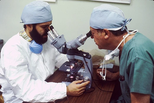
Electron microscope
Uses electron beams for much higher resolution than light microscopes.

An electron microscope is a microscope that uses a beam of electrons as a source of illumination, employing electron optics analogous to the glass lenses of an optical light microscope to control the electron beam. Because the wavelength of an electron can be more than 100,000 times smaller than that of visible light, electron microscopes achieve a much higher resolution of about 0.1 nm, compared to about 200 nm for light microscopes. Types include the transmission electron microscope (TEM), scanning electron microscope (SEM), scanning transmission electron microscope (STEM), electron microprobe, low-energy electron microscope (LEEM), and photoemission electron microscope (PEEM).
- Invented by
- Max Knoll and Ernst Ruska (1931); also Reinhold Rüdenberg (patents filed 1932)
- First exceeded light microscope resoluti
- 1933
- First commercial electron microscope
- 1938 (Siemens)
- Resolution
- about 0.1 nm (modern TEMs below 0.5 angstrom)
- Key developers
- Ernst Ruska, Max Knoll, Reinhold Rüdenberg, Manfred von Ardenne, Albert Crewe
Lore & Background
The groundwork for electron optics included Hertz's 1883 cathode-ray tube with electrostatic and magnetic deflection, Emil Wiechert's 1899 focusing of electrons by an axial magnetic field, Arthur Wehnelt's 1905 improved oxide-coated cathodes, and Hans Busch's 1926 development of the electromagnetic lens. In 1928, Leó Szilárd tried to convince Dennis Gabor to build an electron microscope, for which Szilárd had filed a patent. The issue of who invented the transmission electron microscope remains controversial. In 1928, Adolf Matthias appointed Max Knoll to lead a team at the Technische Hochschule in Charlottenburg; the team included Ernst Ruska. In 1931, Knoll and Ruska generated magnified images of mesh grids using two magnetic lenses, creating the first electron microscope. Reinhold Rüdenberg at Siemens-Schuckert independently filed U.S. patents in 1932, claiming earlier work, but it is not clear when he had a working instrument.
Reader's Guide
Electron microscopes revolutionized imaging by enabling visualization of structures far smaller than possible with light microscopes. The transmission electron microscope (TEM) was the original form, using high-voltage electrons transmitted through thin specimens. The scanning electron microscope (SEM) probes specimens with a focused beam to produce images of surface topography and composition. The scanning transmission electron microscope (STEM) combines features of both. Key milestones include the 1933 instrument exceeding light microscope resolution, the 1937 development of the scanning electron microscope by Manfred von Ardenne, and the 1965 introduction of the scanning transmission electron microscope with a field emission source by Albert Crewe. By the early 1980s, improvements enabled atomic-scale imaging. The 2000s saw advancements in aberration-corrected electron microscopy, significantly improving resolution and clarity. These instruments remain essential in materials science, biology, and nanotechnology.
Did You Know?
- The first electron microscope was built in 1931 by Max Knoll and Ernst Ruska.
- Reinhold Rüdenberg filed U.S. patents for the electron microscope in 1932, but it is not clear when he had a working instrument.
- The first commercial electron microscope was produced by Siemens in 1938.
More in Pathology 1-24
Spotted an error? Know more?
Reader corrections go straight into our review queue. Suggest an edit · How this site is sourced