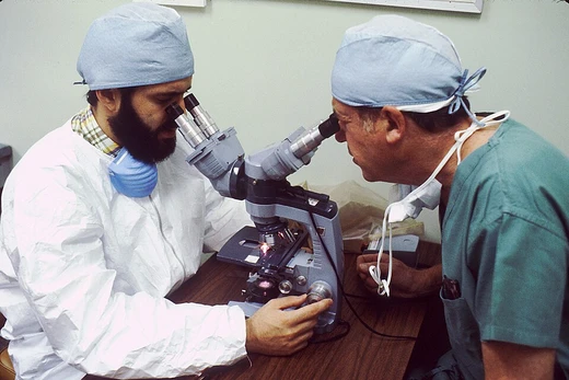
Blood smear
A stained blood layer for microscopic cell analysis.

A blood smear, also known as a peripheral blood smear or blood film, is a thin layer of blood spread on a glass microscope slide, stained to allow microscopic examination of blood cells. It is a fundamental tool in the investigation of hematological disorders and for detecting blood parasites such as those causing malaria and filariasis.
Quick Facts
- Medlineplus
- 003665
Facts from the source article.
Lore & Background
The blood smear is prepared by placing a drop of blood on one end of a slide and using a spreader slide to disperse it, creating a monolayer in the 'feathered edge' where cells are spaced for counting. After air drying, the blood is fixed with methanol to preserve cellular detail, then stained—commonly with Romanowsky stains such as Wright's, Giemsa, or Diff-Quik—to distinguish cell types. The monolayer is viewed under magnification up to 1000 times to characterize cell morphology.
Reader's Guide
Blood smear examination is typically performed alongside a complete blood count to investigate abnormal results or confirm flagged findings. It is essential for determining the cause of anemia by assessing red blood cell shape, size, and coloration, revealing disorders like iron deficiency anemia, sickle cell anemia, megaloblastic anemia, and microangiopathic hemolytic anemia. The manual white blood cell differential from a smear can detect abnormal proportions (e.g., neutrophilia, eosinophilia) and abnormal cells such as circulating blast cells in acute leukemia, as well as qualitative abnormalities like toxic granulation. Although automated analyzers provide differentials, they have limited ability to identify immature and abnormal cells, so manual smear examination remains frequently indicated. For parasitic infections like malaria and babesiosis, blood smear microscopy is the preferred diagnostic method. In malaria diagnosis, thin smears allow species identification, while thick smears are about eleven times more sensitive for detecting low-level infections, though species distinction is more difficult. Proper technique is critical: delays in smear preparation can cause artifacts, such as gametocyte division mimicking Borrelia or acid-induced shrinkage mimicking P. malariae. Staining pH must be 7.2 for malaria films to visualize Schüffner's and James' dots.
Did You Know?
- The monolayer is found in the 'feathered edge' created by the spreader slide as it draws the blood forward.
- Thick smears are about eleven times more sensitive than thin smears for detecting malaria parasites.
- If blood cools to room temperature, male gametocytes can divide and release exflagellated microgametes that may be mistaken for other artifacts or parasites.
- Malaria blood films are typically stained at a pH around 7.0–7.2; within this range, Schüffner's dots and James' dots remain visible.
The Art of Preparation
A blood smear begins with a simple drop of blood placed at one end of a glass slide. A spreader slide is then drawn across, pulling the blood into a thin, even layer. The critical target is the feathered edge — the tapered region where cells separate into a monolayer, spaced just far enough apart for individual counting and morphological assessment. Once the slide air-dries, a brief dip in methanol locks the cells in place, a step that is non-negotiable for preserving fine cellular detail during staining. The staining phase typically employs Romanowsky-family dyes: Wright's stain, Giemsa stain, or the rapid Diff-Quik, with Wright-Giemsa combinations also popular in practice. These colorants render white blood cells, red blood cells, and platelets visually distinct, making abnormalities in each lineage apparent. Hematopathologists reaching beyond routine work may turn to additional specialized stains to sharpen a differential diagnosis. The finished slide is then examined under a microscope at magnifications reaching 1000×, where each cell's shape, size, and internal architecture is carefully characterized and recorded.
Hematological Detective Work
In the clinical laboratory, a blood smear is rarely examined in isolation; it is most often paired with a complete blood count to probe abnormal values or to verify results that an automated analyzer has flagged as questionable. The red-cell compartment tells a rich story: the shape, size, and coloration of erythrocytes can point toward specific anemias, from the small pale cells of iron deficiency to the crescent shapes of sickle cell disease, the oversized forms of megaloblastic anemia, or the fragmented cells of microangiopathic hemolytic anemia. On the white-cell side, a manual differential reveals the proportions of each leukocyte type, flagging shifts such as neutrophilia or eosinophilia. Equally important is the detection of abnormal populations — circulating blast cells that betray acute leukemia, or qualitative changes like toxic granulation. Modern automated analyzers can generate a differential, yet they struggle with immature and atypical cells, which is why the manual smear remains a frequent and essential follow-up.
The Malaria Microscope
Microscopic examination of a blood smear remains the gold standard for diagnosing malaria, because each of the four major Plasmodium species carries distinguishing morphological features. Two smear types work in tandem: thin smears preserve parasite architecture for confident species identification, while thick smears screen roughly eleven times the blood volume, making low-level infections easier to detect at the cost of considerable distortion. A persistent challenge is that early trophozoites — the ring forms — look identical across all four species, so no single ring can assign a species; the microscopist must examine several trophozoites before concluding. Timing is another critical variable. If a sample cools before the smear is made, male gametocytes divide and release long, sinuous microgametes that can be mistaken for Borrelia. Warmer storage causes schizonts to rupture, with merozoites mimicking the accolé form of P. falciparum. Prolonged EDTA exposure shrinks parasitized cells in P. vivax or P. ovale, producing a rolled-up appearance resembling P. malariae. Staining pH also matters: malaria films require pH 7.2 rather than the 6.8 used in routine hematology, or Schüffner's dots and James' dots will not develop.
Beyond the Routine: Parasites, Pathogens & Alternatives
While malaria commands the most attention, the blood smear serves as a frontline screen for a broader cast of infectious agents. Filariasis and babesiosis are among the parasitic conditions for which microscopic examination of the blood film is the preferred diagnostic route. In rarer but dramatic cases, bacteria themselves may be visible on the smear in patients suffering from severe sepsis, offering a glimpse of the organism directly in the patient's circulation. For laboratories that lack trained microscopists or appropriate microscopy expertise, immunochromatographic capture procedures — commonly known as rapid diagnostic tests or malaria antigen detection tests — provide a nonmicroscopic alternative. These point-of-care options can deliver a useful signal when the full microscopy pipeline is unavailable, though they do not replicate the species-level resolution or the morphological detail that a well-prepared, properly stained smear offers under the lens.
Frequently Asked Questions
Who is Blood smear?
A blood smear (also called a peripheral blood smear or blood film) is a thin, stained layer of blood spread on a glass microscope slide so that individual cells can be inspected under magnification. It is a cornerstone technique in hematology and clinical pathology.
What are Blood smear's powers/role?
Its main job is letting a pathologist visually examine red cells, white cells, and platelets to diagnose anemia, leukemia, and parasitic infections like malaria or filariasis. At up to 1000× magnification it reveals morphological clues—cell shape, inclusions, abnormal nuclei—that automated counters often miss.
How is Blood smear prepared?
A small drop of blood is spread across a clean slide, air-dried, fixed in methanol, and then stained with a Romanowsky-type dye such as Wright's, Giemsa, or Diff-Quik. The Wright-Giemsa combination is a popular choice for bringing out fine nuclear and cytoplasmic detail.
Why is Blood smear important?
It remains a fast, inexpensive, first-line test for investigating hematological disorders and detecting blood-borne parasites. Because a trained eye can directly judge cell morphology, it stays indispensable even in the era of fully automated analyzers.
What are Blood smear's signature tools?
Its go-to stains are Wright's stain, Giemsa stain, Diff-Quik, and the Wright-Giemsa blend, all Romanowsky-type dyes that differentially color nuclei versus cytoplasm. Examination is typically done with an oil-immersion objective at magnifications up to 1000×.
More in Pathology 1-24
Spotted an error? Know more?
Reader corrections go straight into our review queue. Suggest an edit · How this site is sourced