Rectus abdominis muscle
Paired abdominal muscle responsible for trunk flexion and posture.

Russ Anderson from London, UK · CC BY 2.0
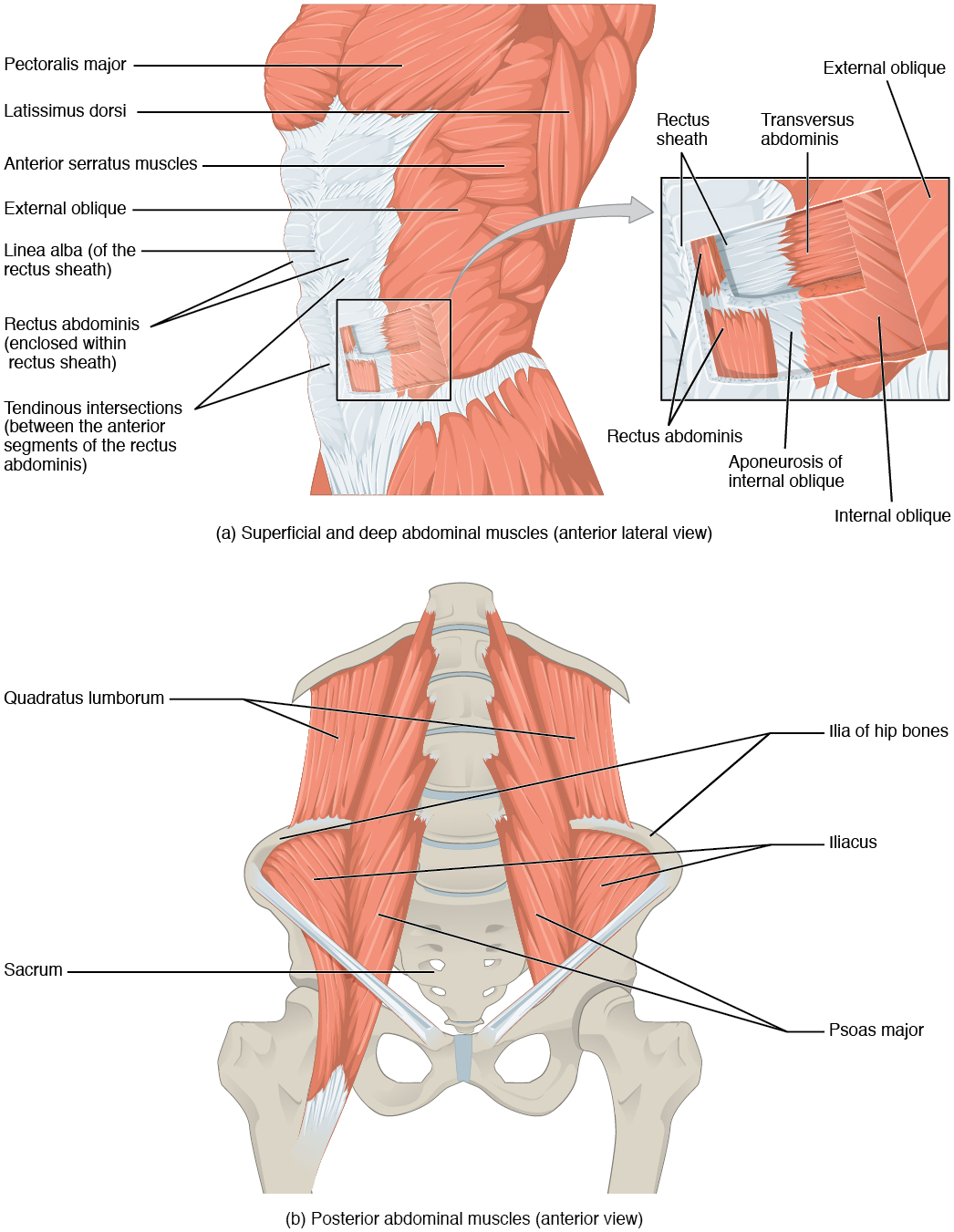
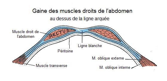
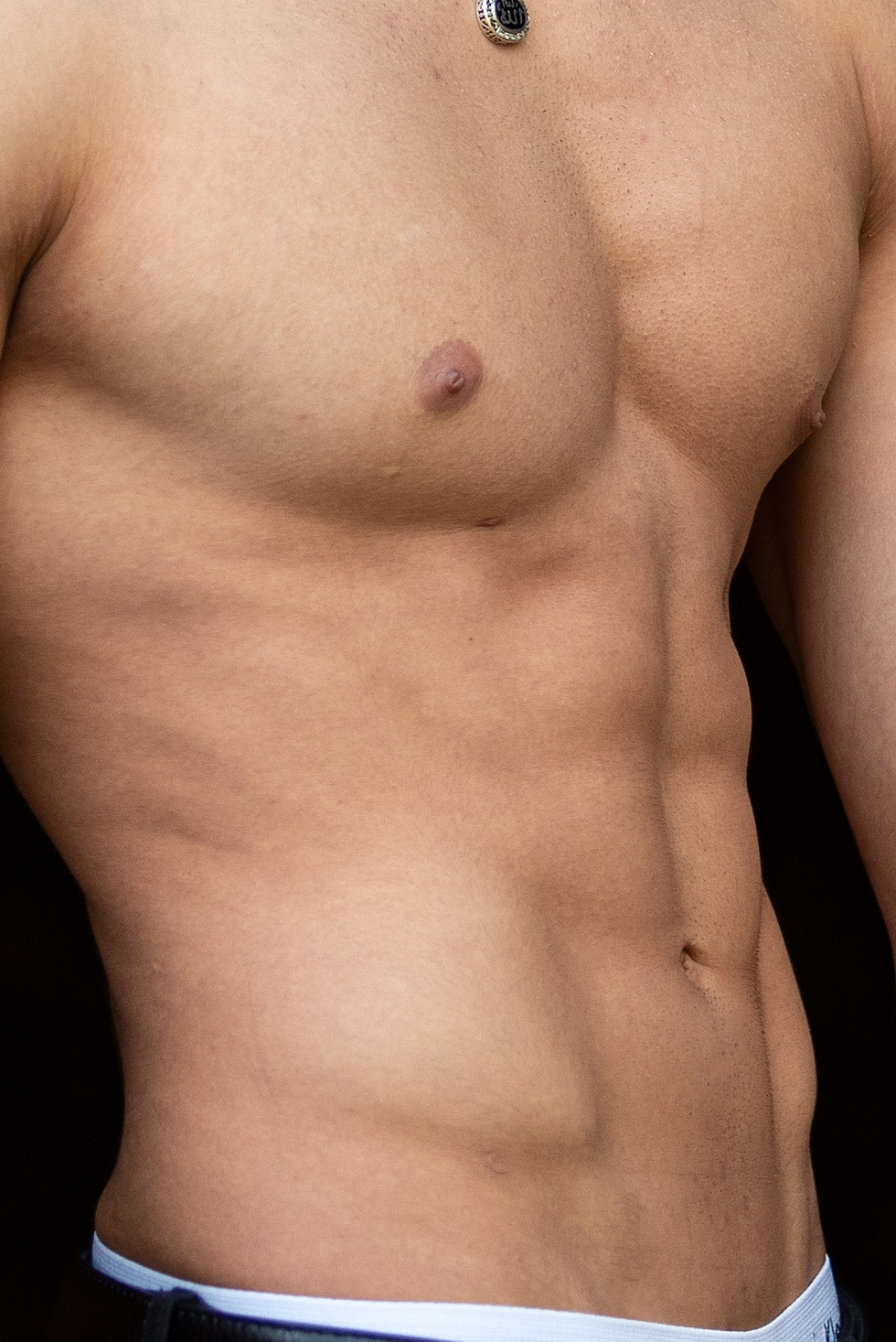
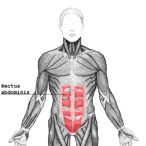
The rectus abdominis, often called the abs or six-pack, is a pair of segmented skeletal muscles running down the front of the abdomen. The two muscles are divided down the middle by a dense connective tissue band called the linea alba, and each side is bordered laterally by the linea semilunaris. Each muscle stretches from the pubic symphysis, pubic crest, and pubic tubercle at the bottom up to the xiphoid process and the costal cartilages of the fifth through seventh ribs at the top. It sits inside the rectus sheath, which is formed by the aponeuroses of the lateral abdominal muscles. Bands of connective tissue called tendinous intersections cross each rectus abdominis, breaking it into distinct muscle bellies.
The muscle itself is long and flat, running the entire length of the front of the abdomen. The tendinous intersections subdivide each side into smaller bellies, and when the muscle tenses, it expands between these intersections. The upper part attaches mainly to the cartilage of the fifth rib, with some fibers also reaching the front end of that rib.
In terms of size, the rectus abdominis is typically about 10 mm thick, though some athletes may have one up to 20 mm thick. Its volume is roughly 300 cm³ in non-active people and around 500 cm³ in athletes.
Blood supply comes from several sources. The inferior epigastric artery and vein run upward along the back of the muscle, entering the rectus fascia at the arcuate line to serve the lower portion. The superior epigastric artery, a terminal branch of the internal thoracic artery, supplies the upper part. Additionally, the lower six intercostal arteries provide small segmental contributions.
The muscles are innervated by the thoraco-abdominal nerves, which are continuations of the T7–T11 intercostal nerves and pierce the front layer of the rectus sheath. Sensory supply comes from the seventh through twelfth thoracic nerves.
A variation called the sternalis muscle may be a variant of the pectoralis major or the rectus abdominis. Some fibers occasionally connect with the costoxiphoid ligaments and the side of the xiphoid process.
Functionally, the rectus abdominis is a key postural muscle. It flexes the lumbar spine, as in a crunch, bringing the rib cage toward the pelvis when the pelvis is fixed, or pulling the pelvis toward the rib cage (posterior pelvic tilt) when the rib cage is fixed, as in a leg-hip raise. Both can move together when neither is fixed. It also assists with breathing, especially during forceful exhalation after exercise or in conditions like emphysema, and helps keep internal organs in place while generating intra-abdominal pressure during heavy lifting, forceful defecation, or childbirth.
Clinically, an abdominal muscle strain, or pulled abdominal muscle, involves tearing of muscle fibers when the muscle is stretched too far. Most strains cause microscopic tears, but severe injuries can rupture the muscle from its attachment. A rectus sheath hematoma is a blood collection within the rectus sheath, causing abdominal pain with or without a mass. It can result from rupture of the epigastric artery or a muscular tear, and causes include anticoagulation, coughing, pregnancy, abdominal surgery, and trauma. With an aging population and widespread use of anticoagulants, this condition is becoming more common and serious. On abdominal exam, a person may show a positive Carnett's sign. Most hematomas resolve without treatment, though it may take months.
In other vertebrates, the rectus abdominis is similar, but the number of tendinous intersections differs from that in humans.
- field
- Anatomy
- known_for
- Flexing the lumbar spine, postural support, and creating intra-abdominal pressure
- typical_thickness
- Around 10 mm (up to 20 mm in athletes)
Lore & Background
The rectus abdominis is a long, flat muscle contained in the rectus sheath, formed by aponeuroses of the lateral abdominal muscles. It is traversed by tendinous intersections that subdivide it into distinct muscle bellies, and tensing causes expansion between these intersections. The muscle has multiple blood supplies: the inferior epigastric artery and vein serve the lower part, the superior epigastric artery supplies the upper portion, and the lower six intercostal arteries provide segmental contributions. Innervation comes from thoraco-abdominal nerves (T7–T11 intercostal nerves), with sensory supply from the 7–12 thoracic nerves.
Reader's Guide
The rectus abdominis is an important postural muscle that flexes the lumbar spine, bringing the rib cage toward the pelvis or vice versa, and assists in forceful exhalation and creating intra-abdominal pressure during activities like heavy lifting, defecation, or childbirth. Clinically, it can be affected by abdominal muscle strain (pulled muscle) or rectus sheath hematoma, an accumulation of blood often caused by rupture of the epigastric artery or muscular tear, with causes including anticoagulation, coughing, pregnancy, abdominal surgery, and trauma. The muscle is similar in most vertebrates, though the number of tendinous intersections varies in animals. Its role in core stability and common injuries makes it a key focus in sports medicine and general health.
Did You Know?
- The rectus abdominis is separated at the midline by a band of dense connective tissue called the linea alba.
- It is typically around 10 mm thick, but some athletes can have a rectus up to 20 mm thick.
- The muscle is innervated by thoraco-abdominal nerves, continuations of the T7–T11 intercostal nerves.
- A rectus sheath hematoma may be caused by rupture of the epigastric artery or by a muscular tear.
Regional Anatomy and Terminology
The human lower limb is a complex structure comprising thirty bones in each leg, with the femur, tibia, and fibula serving as the principal long bones. Anatomically, the limb is divided into distinct regions: the thigh spans from the hip to the knee, while the segment between the knee and the ankle is designated the lower leg, shank, or crus. Within the lower leg, the anterior surface is called the shin (formed by the tibia and the smaller fibula), and the posterior surface is the calf. The patella, the body's largest sesamoid bone, sits in front of the knee joint. It is worth noting that anatomists reserve the word "leg" exclusively for the crus region, whereas the broader term "lower limb" or "lower extremity" encompasses the entire structure from hip to foot. Most of the skeletal framework presents palpable bony landmarks, though the hip joint and the femoral shaft remain hidden beneath soft tissue.
Bipedal Adaptation and Proportional Evolution
Evolution shaped the human lower limb into a mechanism finely tuned for efficient upright walking, a specialization that distinguishes us from other primates. While chimpanzees and orangutans can briefly adopt a vertical posture, they expend considerable energy doing so, whereas humans walk upright with remarkable economy. This adaptation reshaped the entire body: the center of gravity shifted, internal organs were reorganized, and the trunk's biomechanics were restructured. The double S-curve of the vertebral column now functions as a shock absorber, channeling trunk weight down through the feet. Leg length relative to the trunk tells a striking story of evolutionary divergence: in orangutans the ratio is 111 percent, in chimpanzees 128 percent, and in humans a full 171 percent. Muscles such as the gluteals, knee extensors, and calf musculature were all remodeled for this task. Sex-based differences also exist, with females tending toward greater hip anteversion and tibiofemoral angles, while males typically possess longer femoral and tibial segments.
Skeletal Alignment and the Mechanical Axis
In a normally aligned leg, the major joints form a straight mechanical line known as the Mikulicz line, running from the femoral head through the intercondylar eminence of the tibia to the center of the ankle mortise. In the tibial shaft the mechanical and anatomical axes coincide, but in the femoral shaft they diverge by approximately six degrees, producing a femorotibial angle of 174 degrees. A leg is considered straight when, with the feet together, both the medial malleoli and the medial condyles of the knees make contact. Deviations from this norm create clinical conditions: genu varum occurs when the knee center falls lateral to the axis (intermalleolar distance exceeding three centimeters), while genu valgum arises when it falls medial (intercondylar distance exceeding five centimeters). Both impose uneven joint loading. The collodiaphysial angle between the femoral neck and shaft also shifts with age, declining from roughly 150 degrees at birth to 120 degrees in old age, with pathological extremes producing coxa vara or coxa valga.
Hip Musculature and Multi-Axis Joint Mechanics
The muscles surrounding the hip joint can be organized in several ways: by their innervation source (ventral versus dorsal plexus divisions), by their developmental insertion pattern (a two-layered posterior group and an anterior group), or by their functional role as extensors, flexors, adductors, or abductors. Because many of these muscles span extensive origin and insertion areas, a single muscle often participates in several distinct movements, and some even influence the knee or vertebral joints. The hip itself permits motion along multiple axes: lateral and medial rotation occur along the limb's longitudinal axis, flexion and extension swing along a transverse axis, and abduction with adduction rotate about a sagittal axis. A key example is the iliopsoas complex, a group of two or three muscles sharing a common insertion on the lesser trochanter of the femur, with the psoas major originating from the lowest thoracic vertebra and running along the lumbar spine before descending into the pelvis.
Gallery


Frequently Asked Questions
Who is the Rectus abdominis muscle?
It is a paired, vertically oriented skeletal muscle running along the front of the abdomen, anchored at the pubic bone below and reaching up to the xiphoid process and the costal cartilages of ribs 5–7. Fans affectionately nickname it the 'six-pack' because tendinous intersections divide each side into distinct segments.
What are the Rectus abdominis muscle's powers and role?
Its core abilities include flexing the lumbar spine (think a crunch), maintaining upright postural support, and generating the intra-abdominal pressure needed for coughing, lifting, and straining. It works as a stabilizer for the entire trunk during virtually every movement.
How does the Rectus abdominis muscle's story end?
Inferiorly, the muscle tapers into a broad aponeurosis that inserts onto the pubic symphysis, pubic crest, and pubic tubercle, marking the final stop of its bony journey. Superiorly, its fibers blend into the xiphoid process and the 5th–7th rib costal cartilages, completing its full vertical span.
Why is the Rectus abdominis muscle important to the body?
It is a primary core stabilizer that keeps the spine and pelvis aligned during standing, walking, and lifting. It also contributes the intra-abdominal pressure essential for breathing, defecation, and the pushing phase of childbirth.
How thick is the Rectus abdominis muscle, and does it vary between people?
In a typical adult the muscle measures roughly 10 mm thick, though dedicated athletes can develop it to around 20 mm. The midline gap between the two halves is the linea alba, which is what creates the characteristic segmented, paired appearance fans recognize.
More in Musculoskeletal System And Orthopedics 1-17
Spotted an error? Know more?
This is a living reference — every entry is fact-audited, and reader corrections feed straight into our audit queue. Suggest an edit · See this site's audit record