Pectoralis major
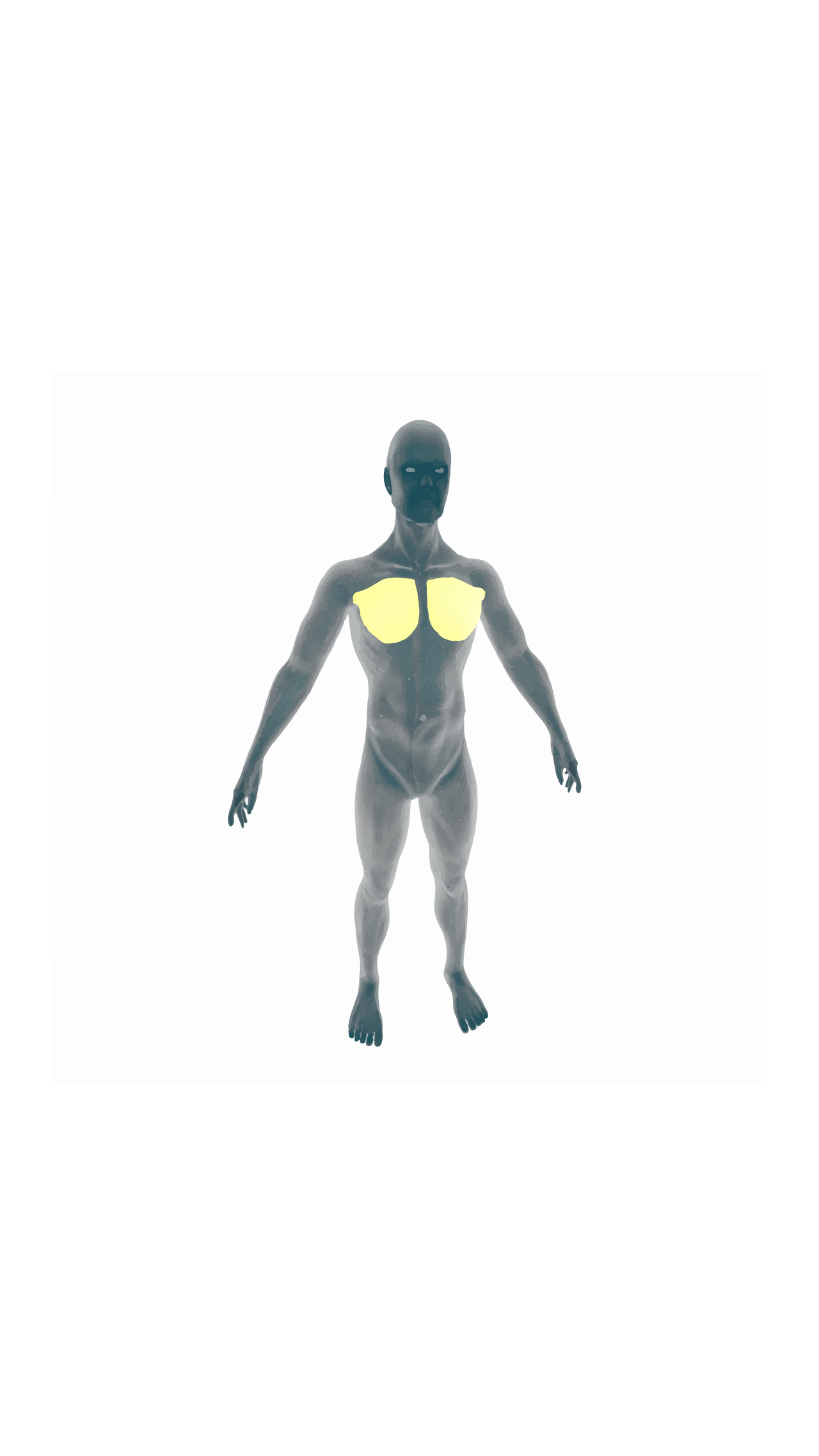
Thick, fan-shaped muscle of the human chest.

DataBase Center for Life Science (DBCLS) · CC BY 4.0
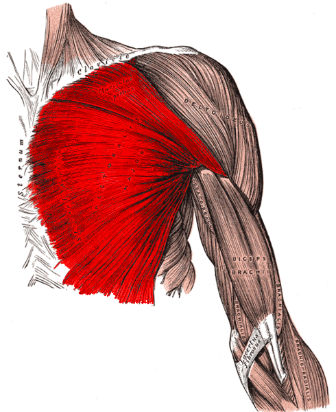
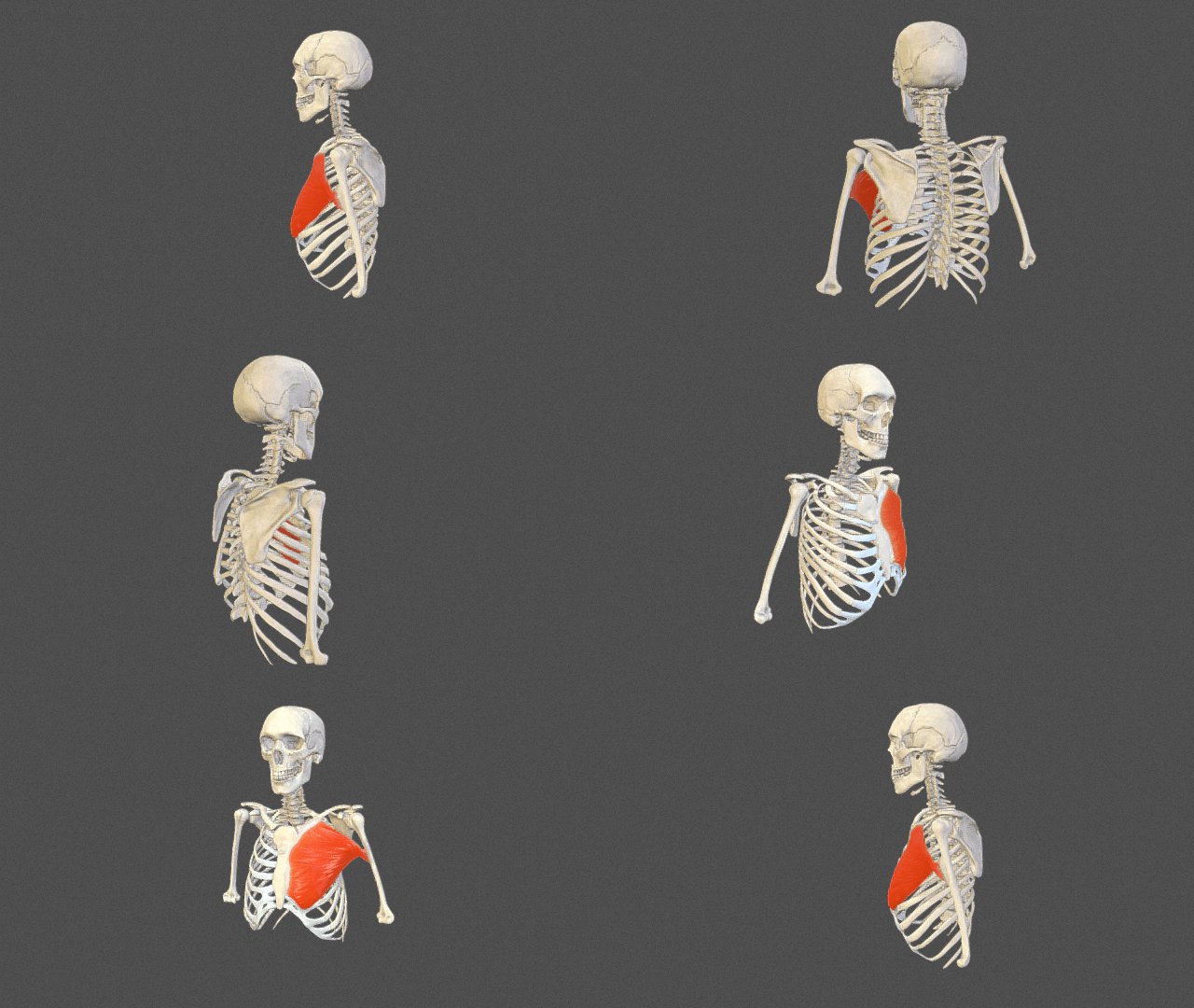
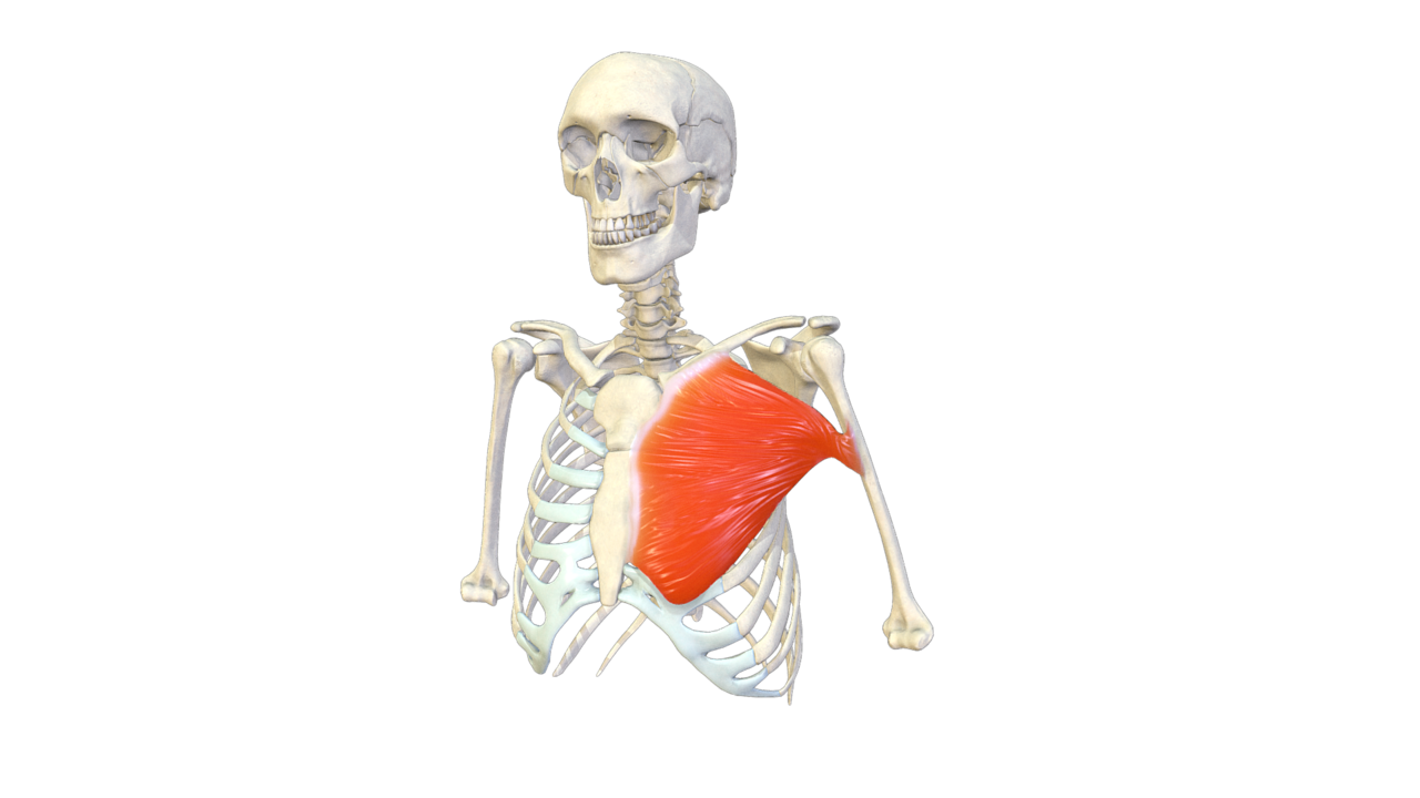
The pectoralis major is a thick, fan-shaped muscle in the chest, its name coming from the Latin word for "breast." It forms the bulk of the chest musculature, sitting just beneath the breast tissue, with the pectoralis minor lying deeper underneath. This muscle originates from the clavicle, the sternum, the costal cartilages of the true ribs, and the aponeurosis of the external oblique abdominal muscle. It inserts into the lateral lip of the bicipital groove on the humerus. Two nerves supply it: the medial and lateral pectoral nerves. Its main jobs are flexing, adducting, and internally rotating the upper arm bone. Commonly called "pecs," the pectoral muscle, or chest muscle, it is the largest and most superficial muscle in the chest area.
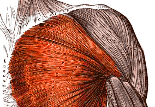
The muscle's fibers start from the front surface of the sternal half of the clavicle, the full width of the sternum's front half down to the sixth or seventh rib, the cartilages of all true ribs (often except the first or seventh), and the abdominal external oblique's aponeurosis. From these broad origins, the fibers converge toward their insertion. Clavicular fibers run obliquely downward and outward, often separated from the rest by a small gap. Fibers from the lower sternum and lower true rib cartilages run upward and outward, while middle fibers go horizontally. All end in a flat tendon about 5 cm wide, inserting into the humerus's bicipital groove. This tendon has two laminae, front and back, usually fused below. The thicker front lamina receives clavicular and upper sternal fibers, with the most lateral clavicular fibers inserting at its top and upper sternal fibers going to its lower part, which extends down to and joins the deltoid tendon. The back lamina gets most of the sternal portion and deep fibers from the costal cartilages. These deep fibers, especially from lower costal cartilages, ascend higher on the humerus, twisting behind the superficial fibers, so the tendon appears twisted. The back lamina reaches higher on the humerus than the front and gives an expansion covering the intertubercular groove, blending with the shoulder joint capsule. From the deepest fibers at insertion, an expansion lines the intertubercular groove, and from the tendon's lower border, a third expansion goes down to the arm's fascia.
The muscle gets dual motor innervation from the medial and lateral pectoral nerves. The sternal head is innervated by C7, C8, and T1 nerve roots via the lower trunk of the brachial plexus and the medial pectoral nerve. The clavicular head gets innervation from C5 and C6 nerve roots via the upper trunk and lateral cord of the brachial plexus, which gives off the lateral pectoral nerve, distributed over the muscle's deep surface. Sensory feedback follows the reverse path, returning via first-order neurons to spinal nerves at C5, C6, C8, and T1 through posterior rami. After synapsing in the spinal cord's posterior horn, sensory information about movement, proprioception, and pressure travels via a second-order neuron in the dorsal column medial lemniscus tract to the medulla. There, fibers decussate to form the medial lemniscus, carrying sensory information to the thalamus. The thalamus diverts some info to the cerebellum and basal nuclei for motor feedback, while some ascends directly to the postcentral gyrus of the parietal lobe via third-order neurons. Sensory info for this muscle is processed in the superior part of the sensory homunculus, near the longitudinal fissure. Electromyography suggests it has at least six independently coordinated muscle fiber groups.
Common variations include greater or lesser attachment to the ribs and sternum, varying size or absence of the abdominal part, separation of sternocostal and clavicular parts, fusion of the clavicular part with the deltoid, and decussation in front of the sternum. Deficiency or absence of the sternocostal part is more common than absence of the clavicular part. Poland syndrome is a rare congenital condition where the whole muscle is missing, often on one side, sometimes with breast absence in females. The sternalis muscle may be a variant of the pectoralis major or rectus abdominis. The chondroepitrochlearis is an uncommon variation, an atypical musculotendinous structure arising from the pectoralis major and attaching to the humerus's medial epicondyle.
The pectoralis major has four primary actions for shoulder joint movement: flexion of the humerus, adduction, internal rotation, and—though not listed in the source—the source text cuts off before completing the fourth action. Based on the provided facts, its known functions are flexion, adduction, and internal rotation of the humerus.
- Origin
- Anterior surface of sternal half of clavicle, anterior surface of sternum, cartilages of true ribs, aponeurosis of abdominal external oblique muscle
- Insertion
- Lateral lip of bicipital groove (intertubercular sulcus) of humerus
- Nerve supply
- Medial pectoral nerve and lateral pectoral nerve
- Actions
- Flexion, adduction, and internal rotation of humerus
- Blood supply
- Not specified in source
Lore & Background
The pectoralis major arises from the anterior surface of the sternal half of the clavicle, from the breadth of the half of the anterior surface of the sternum as low down as the attachment of the cartilage of the sixth or seventh rib, from the cartilages of all the true ribs (with the exception, frequently, of the first or seventh), and from the aponeurosis of the abdominal external oblique muscle. From this extensive origin, the fibers converge toward their insertion; those from the clavicle pass obliquely downward and outward, those from the lower part of the sternum and cartilages of the lower true ribs run upward and laterally, while the middle fibers pass horizontally. They all end in a flat tendon about 5 cm in breadth, inserted into the lateral lip of the bicipital groove. The tendon consists of two laminae, placed one in front of the other, and usually blended together below.
Reader's Guide
The pectoralis major is significant as the largest and most superficial muscle in the chest area, colloquially referred to as 'pecs', 'pectoral muscle', or 'chest muscle'. Its primary functions—flexion, adduction, and internal rotation of the humerus—are essential for movements such as throwing underhand, flapping the arms, and arm-wrestling. The muscle receives dual motor innervation from the medial and lateral pectoral nerves, with the clavicular head innervated by C5 and C6 nerve roots and the sternal head by C7, C8, and T1. Electromyography suggests it consists of at least six groups of muscle fibres that can be independently coordinated by the central nervous system. Clinically, tears of the pectoralis major are rare and typically affect male athletes in contact sports and weight-lifting, particularly during bench press. High-grade partial or full-thickness tears warrant surgical repair if function is to be preserved. Poland syndrome is a rare congenital condition in which the whole muscle is missing, most commonly on one side of the body.
Did You Know?
- The pectoralis major receives double motor innervation from the medial pectoral nerve and the lateral pectoral nerve.
- The tendon of the pectoralis major consists of two laminae, placed one in front of the other, and usually blended together below.
- Poland syndrome is a rare congenital condition in which the whole muscle is missing, most commonly on one side of the body.
- The chondroepitrochlearis is an uncommon muscular variation that arises from the pectoralis major and attaches to the medial epicondyle of the humerus.
Anatomy & Structural Organization
The serratus anterior derives its name from two Latin roots: serrare, meaning to saw, a nod to its jagged, tooth-like contour, and anterior, indicating its position on the front of the body. This chest muscle takes its origin from the lateral surfaces of the upper nine ribs, though it actually produces ten distinct muscle slips because two separate slips arise from the second rib. On the other end, the fibers converge and attach along the entire medial border of the scapula, spanning from the superior angle down to the inferior angle. Based on where they insert, the muscle is conventionally divided into three functional segments: a superior portion attaching near the top of the scapula, an intermediate portion along the mid-border, and an inferior portion near the bottom angle. The long thoracic nerve, a branch of the brachial plexus, provides all motor innervation and runs inferiorly across the muscle's surface. Anatomically, the serratus anterior sits deep to the subscapularis, with a bursa separating the two, while another bursa interposes it from the underlying rib.
Biomechanical Roles & Functional Versatility
The primary action of the serratus anterior is to draw the scapula forward and around the rib cage, a movement called protraction that is essential for raising the arm ahead of the body. In this role it opposes the rhomboids, which pull the scapula medially. Yet the relationship is not purely antagonistic: when the superior and inferior portions contract together, they press the scapula firmly against the thoracic wall, working in concert with the rhomboids as synergists. The inferior segment additionally tugs the lower scapular angle laterally and forward, rotating the bone so that full overhead elevation of the arm becomes possible. In partnership with the upper and lower fibers of the trapezius, the serratus anterior sustains the upward rotation needed for lifting objects above the head. Beyond the shoulder girdle, all three segments can elevate the ribs when the shoulder is held fixed, thereby contributing to the mechanics of respiration. Because of its dominant role in scapular protraction during a punching motion, the muscle has earned the informal nicknames big swing muscle and boxer's muscle.
Clinical Vulnerability & Winged Scapula
The long thoracic nerve, the sole motor supply to the serratus anterior, courses inferiorly directly on the surface of the muscle, making it particularly exposed to iatrogenic injury. During axillary lymph node dissection, commonly performed as part of breast cancer surgery, this nerve is at significant risk of being cut or stretched. When the nerve is damaged, the serratus anterior loses its ability to hold the medial border of the scapula against the rib cage. The result is a condition known as winged scapula, in which the inner edge of the shoulder blade protrudes posteriorly and becomes visibly prominent, especially when the patient pushes against a wall or raises the arms. Nerve injury of this type is the most frequently cited cause of the deformity. The condition is also referenced under the broader label of backpack palsy, a colloquial term for thoracic long nerve compression from heavy shoulder straps. Targeted rehabilitation exercises for the serratus anterior are prescribed to restore scapular stability and reduce the cosmetic and functional impact of the winging.
Evolutionary Origins & Comparative Anatomy
Within the broader classification of shoulder muscles, the serratus anterior belongs to the axioscapular group, alongside the rhomboid major, rhomboid minor, levator scapulae, and trapezius. While the trapezius evolved independently, the remaining four trace their origins to the first eight or ten ribs and the transverse processes of cervical vertebrae. In early tetrapods, the group's principal job was controlling the scapula's vertebral border; fibers for dorsal motion became the rhomboids, those for ventral motion gave rise to the serratus anterior, and those for cranial motion evolved into the levator scapulae. Over time, the serratus anterior underwent three notable changes: its distal and proximal fibers grouped more distinctly, its intermediate fibers shrank, and its dominant superior and inferior portions came to anchor on the scapular angles. In primates, the wide thoracic cage and laterally facing glenoid cavity shift medial forces onto the clavicle. In cursorial quadrupeds like horses, the scapula hangs vertically with no clavicle, and the thorax is suspended between the scapulae by the serratus anterior and pectoralis muscles. In climbing species, the serratus anterior must resist reaction forces of a free limb, generating high bending loads on ribs that are consequently more curved and reinforced by the clavicle.
Gallery

Frequently Asked Questions
What is the Pectoralis major?
It is the thick, fan-shaped muscle that makes up most of the visible chest wall in humans, sitting directly beneath the breast tissue. Fans often call it the main event of the pectoral region, with the smaller Pectoralis minor tucked as a supporting layer underneath.
Where does the Pectoralis major attach?
It fans out from the front of the clavicle, the sternum, the cartilages of the true ribs, and the aponeurosis of the external oblique, then converges to anchor on the lateral lip of the bicipital groove on the humerus.
What movements does the Pectoralis major perform?
It drives the arm forward (flexion), pulls it across the body (adduction), and rotates it inward (internal rotation) at the shoulder. These are the same actions you see in a classic push-up or a tight hug.
Which nerves supply the Pectoralis major?
It receives a dual motor supply from both the medial pectoral nerve and the lateral pectoral nerve. This double wiring is one reason it can generate such strong, reliable shoulder movement.
How does the Pectoralis major relate to the Pectoralis minor?
The Pectoralis minor sits directly beneath the Pectoralis major, forming a thinner, deeper layer in the same chest region. Together they make up the pectoral group, but the major is the thick, fan-shaped workhorse while the minor is a slimmer supporting player.
More in Musculoskeletal System And Orthopedics 1-17
Spotted an error? Know more?
This is a living reference — every entry is fact-audited, and reader corrections feed straight into our audit queue. Suggest an edit · See this site's audit record