Stomach
A muscular digestive organ that breaks down food chemically and mechanically.

The stomach is a muscular, hollow organ found in the upper gastrointestinal tract of humans, many other animals, and even some invertebrates. Its ancient Greek name, *gaster*, gives us the medical term "gastric." This dilated organ is a key part of the digestive system, taking over after the cephalic phase—triggered by seeing, smelling, and chewing food—to begin the gastric phase of digestion. Inside, digestive enzymes and gastric acid chemically break down food, and the stomach also helps manage the gut microbiota, which influences both digestion and overall health.
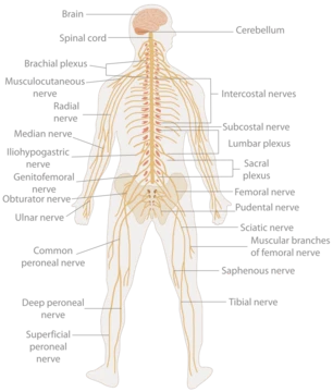
Situated between the esophagus and the small intestine, the stomach sits in the left upper quadrant of the abdominal cavity, with its top against the diaphragm and the pancreas behind it. A large fold of visceral peritoneum called the greater omentum hangs from its greater curvature. Two sphincters keep its contents contained: the lower esophageal sphincter at the junction with the esophagus, and the pyloric sphincter, which controls the release of partially digested food (chyme) into the duodenum, the first part of the small intestine. The stomach is surrounded by parasympathetic and sympathetic nerve networks that regulate both its secretion of digestive juices and the movement of its muscles.
The stomach is distensible, normally expanding to hold about one litre of food, though maximum adult volume ranges from 2 to 4 litres, with extreme cases reaching 15 litres. Its shape changes with distension: empty, it is somewhat J-shaped; partially full, it becomes pear-shaped; in obese individuals, it is more horizontal. A newborn’s stomach holds only about 30 millilitres.
The human stomach has four sections. The cardia, a small region around the esophageal opening, receives food from the esophagus via the cardiac orifice. A cardiac notch marks the start of the greater curvature, and a horizontal line from this notch defines the dome-shaped fundus. The body, or corpus, is the main central region. The pylorus connects to the duodenum at the pyloric sphincter, with the pyloric antrum opening into the body. The cardia begins at the z-line, where the epithelium changes from stratified squamous to columnar, near the lower esophageal sphincter.
The stomach bed—the structures it rests upon—includes the tail of the pancreas, splenic artery, left kidney, left suprarenal gland, transverse colon and its mesocolon, left crus of diaphragm, and left colic flexure. This term was introduced around 1896 but later discredited.
Blood supply comes from the right and left gastric arteries along the lesser curvature, the right and left gastroepiploic arteries along the greater curvature, and 5–7 short gastric arteries from the splenic artery to the fundus and upper greater curvature. Lymphatic drainage occurs via two sets of gastric lymph nodes, which send fluid into the intestinal lymph trunk and then to the cisterna chyli.
The stomach wall, from inner to outer, consists of mucosa, submucosa, muscular layer, subserosa, and serosa. The inner gastric mucosa is a mucous membrane with columnar epithelium, lamina propria, and a thin smooth muscle layer called the muscularis mucosa. Beneath it, the submucosa contains fibrous connective tissue and Meissner's plexus. The muscular layer has three fibre layers—inner oblique, middle circular, and outer longitudinal—arranged at angles to each other. The inner oblique layer is unique to the stomach and creates the churning motion that physically breaks down food. This three-layer structure gives the stomach the thickest muscular wall in the gastrointestinal tract, allowing for maximum peristalsis.
- Location
- Between the esophagus and the small intestine, in the left upper quadrant of the abdominal cavity
- Capacity
- Normally expands to hold about one litre; maximum adult volume 2–4 litres; up to 15 litres in extreme circumstances
- Sections
- Cardia, fundus, body (corpus), and pylorus
- Muscle layers
- Three layers: inner oblique, middle circular, outer longitudinal
- Sphincters
- Lower esophageal sphincter and pyloric sphincter
Lore & Background
The stomach is a muscular, hollow, and highly distensible organ situated in the upper left quadrant of the abdominal cavity, lying between the esophagus and the duodenum. Its shape is not fixed; when empty it is somewhat J-shaped, becoming pyriform (pear-shaped) when partially distended, and more horizontal in obese individuals. In a newborn, the stomach holds only about 30 millilitres, while an adult can normally expand to hold roughly one litre, with a maximum volume between 2 and 4 litres, though volumes up to 15 litres have been observed in extreme cases. The organ is divided into four sections: the cardia, a small region surrounding the esophageal opening where stratified squamous epithelium changes to columnar at the z-line; the fundus, a dome-shaped upper curved part; the body or corpus, the main central region; and the pylorus, which connects to the duodenum via the pyloric sphincter. Two sphincters contain stomach contents: the lower esophageal sphincter at the cardiac region and the pyloric sphincter. The stomach wall, like other parts of the gastrointestinal tract, consists of an inner mucosa (with columnar epithelium, lamina propria, and muscularis mucosa), a submucosa of fibrous connective tissue containing Meissner’s plexus, and a muscular layer unique for having three layers—inner oblique, middle circular, and outer longitudinal—with fibres lying at angles to each other. The stomach is surrounded by parasympathetic and sympathetic nerve plexuses that regulate both its secretory and motor activities. The term “gastric” derives from the Ancient Greek *gaster*.
Reader's Guide
The stomach is a central organ in the digestive system, responsible for the chemical and mechanical breakdown of food. Its unique three-layered muscular wall allows for churning and peristalsis, while gastric glands secrete acid and enzymes that initiate protein digestion. The stomach also regulates gut microbiota, influencing overall health. Its distensibility allows it to accommodate varying volumes, from 30 millilitres in newborns to up to 15 litres in extreme cases. The stomach's four sections—cardia, fundus, body, and pylorus—each have specialized functions, with the pyloric sphincter controlling the release of chyme into the small intestine. The organ's blood supply and lymphatic drainage support its metabolic and immune functions. The stomach's role in digestion and its anatomical complexity make it a key subject in medical study, though the term 'stomach bed' remains disputed.
Did You Know?
- The stomach can normally expand to hold about one litre of food, but maximum adult volume is between 2 and 4 litres, with up to 15 litres observed in extreme circumstances.
- The stomach wall contains three layers of muscle—inner oblique, middle circular, and outer longitudinal—with the inner oblique layer unique to the stomach.
- The pyloric sphincter controls the passage of partially digested food (chyme) from the stomach into the duodenum.
More in Human Anatomy 1-24
Spotted an error? Know more?
This is a living reference — every entry is fact-audited, and reader corrections feed straight into our audit queue. Suggest an edit · See this site's audit record