Eye
Sensory organ that detects light and enables vision.

An eye is a biological organ that detects light and transforms it into electrical signals within neurons, forming part of an organism’s visual system. In more advanced animals, the eye functions as an intricate optical device: it gathers light from the surroundings, controls its brightness with a diaphragm, uses adjustable lenses to focus an image, converts that image into electrical impulses, and sends those signals to the brain via the optic nerve and other neural pathways.
Eyes capable of resolving detail appear in ten distinct structural forms, split into two main categories: compound eyes and non-compound eyes. Compound eyes, common in insects and crustaceans, consist of many tiny visual units. Non-compound eyes, found in mammals including humans, have a single lens that focuses light onto the retina to produce one image. The simplest eyes are pit eyes, which are light-sensitive spots set into a depression; this arrangement narrows the incoming light angle, helping the organism determine where the light is coming from.
Beyond vision, eyes also handle several light-driven responses. In animals with more complex eyes, specialized retinal ganglion cells send signals along the retinohypothalamic tract to the suprachiasmatic nuclei for circadian rhythm regulation, and to the pretectal area for the pupillary light reflex.
Complex eyes can distinguish shapes and colors. Many predators have large areas of binocular vision for depth perception, while prey animals like rabbits and horses often have eyes positioned to maximize their field of view, giving them monocular vision. The first proto-eyes appeared around 600 million years ago, near the time of the Cambrian explosion. The last common ancestor of all animals already had the biochemical tools needed for vision, and since then, more advanced eyes have evolved in 96% of animal species across six of the roughly 35 major phyla. In most vertebrates and some molluscs, light enters the eye and lands on the retina, a light-sensitive layer. Cone cells detect color, and rod cells handle low-light contrast; both convert light into neural signals sent via the optic nerve to the brain. Such eyes are usually spherical, filled with a clear gel called vitreous humour, and have a focusing lens and often an iris. Muscles around the iris adjust the pupil size to control light entry and reduce optical aberrations. In many cephalopods, fish, amphibians, and snakes, the lens shape is fixed, and focusing happens by moving the lens forward or backward, like a camera.
Arthropod compound eyes are built from many simple facets. Depending on their anatomy, these facets can produce either a single pixelated image or multiple images per eye. Each sensor has its own lens and light-sensitive cells. Some eyes contain up to 28,000 such sensors arranged in a hexagonal pattern, giving a full 360° field of view. Compound eyes are especially sensitive to motion. Some arthropods, like many Strepsiptera, have compound eyes with only a few facets, each capable of forming an image; the brain then fuses these separate images into one high-resolution picture. The mantis shrimp possesses the most complex color vision system known, with detailed hyperspectral color vision. Extinct trilobites had unique compound eyes made of clear calcite crystals, unlike the soft eyes of most other arthropods. The number of lenses in a trilobite eye varied widely, from just one to thousands per eye.
Simple eyes, in contrast, have a single lens. Jumping spiders have one large pair of simple eyes with a narrow field of view, plus smaller eyes for peripheral vision. Some insect larvae, such as caterpillars, have simple eyes called stemmata. These usually provide only a rough image, but in sawfly larvae they can resolve details down to 4 degrees of arc, detect polarized light, and increase their sensitivity at night by a factor of 1,000 or more. Ocelli, among the simplest eyes, are found in animals like some snails. They have light-sensitive cells but no lens or image-forming structure, so they can only tell light from dark, helping the animal avoid direct sunlight. In creatures living near deep-sea vents, compound eyes are adapted to detect the infrared light from the hot vents, allowing them to avoid being boiled alive.
There are ten different eye layouts. They can be grouped into "simple eyes," which have one concave photoreceptive surface, and "compound eyes," which have many individual lenses on a convex surface. The term "simple" does not mean less complex or less acute; any eye type can be adapted for nearly any behavior or environment. The main limitation is resolution: compound eyes, due to physics, cannot achieve better than 1° resolution. Superposition eyes are more sensitive than apposition eyes, making them better for dark-dwelling creatures. Eyes also fall into two groups based on photoreceptor cell structure: ciliated (as in vertebrates) or rhabdomeric. These groups are not monophyletic—Cnidaria have ciliated cells, and some gastropods and annelids have both types. Some organisms possess photosensitive cells without forming a true eye.
- prevalence
- Found in about 95-96% of animal species across roughly 30 phyla
Lore & Background
Eyes with resolving power come in several fundamentally different forms, commonly classified into compound eyes and camera-type eyes. Compound eyes, made up of multiple small visual units (ommatidia), are common on insects and crustaceans. Camera-type eyes have a single lens and focus light onto the retina to form a single image, as seen in mammals including humans. The simplest eyes are pit eyes, which are eye-spots set into a pit to reduce the angle of incoming light, allowing the organism to deduce the angle of light. More complex eyes include spherical lens eyes, which incorporate a material with higher refractive index to improve resolution. Camera-type eyes have evolved independently at least 8 to 10 times across various groups, including gastropods, annelids, cephalopods, and chitons.
Reader's Guide
The eye is a fundamental sensory organ that has shaped the evolution of animal behavior and ecology. Its ability to detect light and form images allows organisms to navigate, find food, avoid predators, and communicate. The diversity of eye types—from simple pit eyes to complex camera-like eyes and compound eyes—reflects adaptation to different environments and lifestyles. The mantis shrimp possesses the world's most complex color vision system, with detailed hyperspectral color vision. Eyes also serve non-visual functions, such as regulating circadian rhythms through the retinohypothalamic tract and controlling the pupillary light reflex. The study of eyes has informed technological advances in optics and imaging, as every common method of capturing an optical image occurs in nature, except zoom and Fresnel lenses.
Did You Know?
- The mantis shrimp has the world's most complex colour vision system, with detailed hyperspectral colour vision.
- Trilobites had unique compound eyes with lenses made of clear calcite crystals.
The Layered Architecture of the Body
The human body is not a single undifferentiated mass but a nested hierarchy of biological organization. At the most fundamental level, cells and connective tissue form the building blocks. These assemble into tissues, which in turn compose organs, and organs are grouped into the body's major biological systems. This layered structure means that understanding the whole requires understanding each tier and how it relates to the next. Anatomy itself splits along a similar scale. Gross anatomy—sometimes called macroscopic, topographical, or regional anatomy—concerns structures visible without magnification. Microscopic anatomy, by contrast, demands the aid of microscopes and branches into histology, the study of how tissues are organized, and cytology, the study of individual cells. These anatomical investigations do not exist in isolation. They work hand in hand with human physiology, which examines how structures function, and biochemistry, which probes the chemical processes within living matter. Together, the three form the core basic medical sciences that anchor a student's first year of medical training.
Learning the Body: Education and Practice
The path to mastering human anatomy is both theoretical and deeply hands-on. Students in medicine, dentistry, physiotherapy, nursing, paramedicine, and radiography encounter the subject through a blend of anatomical models, skeletal specimens, textbooks, illustrated diagrams, photographs, classroom lectures, and small-group tutorials. For those pursuing medical or dental degrees, the curriculum adds a particularly visceral component: the practical dissection and close inspection of human cadavers. Microscopic anatomy is reinforced by examining prepared histological slides under a microscope, allowing students to see tissue architecture at a scale invisible to the unaided eye. A thorough working knowledge of anatomy is not optional; it is a professional requirement for every physician, with heightened importance for surgeons and for specialists in diagnostic fields such as histopathology and radiology. The subject is typically delivered in the first year of medical school, and it can be organized either regionally—moving from head and neck to thorax, abdomen, and limbs—or systemically, tracing the nervous, respiratory, circulatory, and other functional systems. The landmark reference Gray's Anatomy has recently shifted from a systems-based layout to a regional one, mirroring the way modern classrooms now structure the material.
Anatomy as an Artistic Discipline
Long before modern imaging technology existed, the human body was one of the great subjects of visual art, and understanding its structure became essential to the craft. Gross anatomy—knowledge of how bones articulate and how muscles deform with movement—underpins the work of anyone drawing, painting, or animating a human figure. Dedicated guides, such as Human Anatomy for Artists: The Elements of Form, exist specifically to help artists render the body with anatomical accuracy. Perhaps the most celebrated intersection of art and anatomy belongs to Leonardo da Vinci. He deliberately dissected and studied the human form to sharpen his paintings and drawings, and in doing so he made genuine advances in anatomical knowledge while simultaneously elevating the representation of the human figure in art. His work demonstrated that the two pursuits—scientific understanding and artistic expression—could feed each other. Because the living organism is inherently complex, anatomy must be organized in a structured way, progressing from the smallest cellular components up through tissues, organs, and finally the relationships among organs. This hierarchical logic gives both the artist and the scientist a shared framework for making sense of the body's intricate design.
Regional Maps and Organ Systems
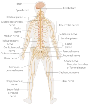
Anatomy can be mapped in two complementary ways. The regional approach divides the body into geographic zones: the head and neck (everything above the thoracic inlet), the upper limb from shoulder to fingertips, the thorax bounded by the thoracic inlet and diaphragm, the abdomen down to the pelvic brim, the back with its vertebrae, sacrum, coccyx, and intervertebral disks, the pelvis and perineum, and the lower limb below the inguinal ligament. Each region houses its own set of internal organs—brain structures, the heart and lungs in the chest, the liver, kidneys, and intestines in the abdomen, and the reproductive organs in the pelvis. The systemic approach, by contrast, groups structures by function. The circulatory system pumps blood through vessels; the digestive system processes food; the endocrine system communicates via hormones from glands like the pituitary, thyroid, and adrenals; the nervous system collects and transmits information through the brain, spinal cord, and nerves; the respiratory system handles breathing; and the urinary system manages fluid and electrolyte balance. The immune, lymphatic, integumentary, musculoskeletal, and reproductive systems round out the full picture, each a network of organs working toward a shared purpose.
Frequently Asked Questions
What is Eye?
Eye is a sensory organ whose core job is to detect incoming light and translate it into electrochemical nerve impulses that the brain can interpret as visual information.
What is Eye's role in the visual system?
It acts as the primary light receptor, capturing photons and converting them into signals carried by neurons so the organism can perceive its surroundings.
How widespread is Eye across the animal kingdom?
This organ is present in roughly 95–96% of all animal species spanning approximately 30 distinct phyla, making it one of the most universally distributed sensory structures in nature.
What are Eye's main evolutionary forms?
Eyes have developed along several fundamentally different evolutionary paths, but they are most commonly grouped into two broad categories: compound eyes and camera-type eyes.
Why is Eye important to an organism?
By enabling vision, it allows organisms to perceive, navigate, and respond to their environment in ways that other sensory channels alone cannot achieve.
More in Human Anatomy 1-24
Spotted an error? Know more?
This is a living reference — every entry is fact-audited, and reader corrections feed straight into our audit queue. Suggest an edit · See this site's audit record