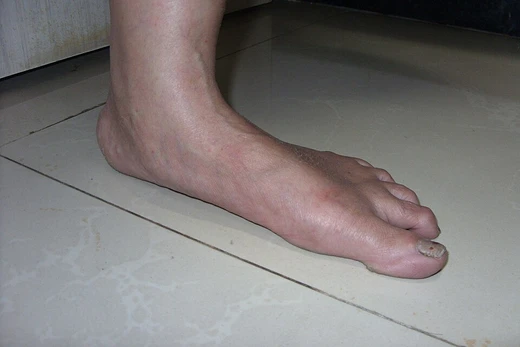
Ainhum
A painful constriction of the fifth toe leading to spontaneous amputation.
Ainhum, also called dactylolysis spontanea, is a condition characterized by a painful constriction at the base of the fifth toe. It frequently leads to bilateral spontaneous autoamputation of the affected toe after several years.
Quick Facts
- Prevalence
- 0.015% to 2.2% of the population
- Sex ratio
- More common in men than in women (2:1)
- Age range
- 20 to 50 years
- Average age
- About thirty-eight
- Youngest recorded patient
- Seven years old
- Pain prevalence
- 78% of cases
- Autoamputation timeframe
- About five years
Facts from the source article.
Did You Know?
- Incisions across the groove are ineffective; excision with z-plasty can relieve pain and prevent autoamputation in Grade I and II lesions.
- Grade III lesions are treated by disarticulating the metatarsophalangeal joint, which relieves pain and yields a useful, stable foot.
- Oral retinoids such as tretinoin and antifibrotic agents like tranilast have been tested for pseudo-ainhum.
Signs and symptoms
The groove begins on the lower and internal side of the base of the fifth toe, typically along the plantar-digital fold, and gradually deepens and becomes more circular. The rate of progression varies, completing a full circle in months or remaining incomplete for years. Both feet are affected in about 75 percent of cases, though usually to different degrees. Only the fifth toe is initially involved, though grooves occasionally appear on the fourth or third toe. The distal part of the toe swells due to lymphatic edema, resembling a small potato. Crusts may form in the groove and become infected with staphylococcus. As the groove deepens, compression of tendons, vessels, and nerves occurs, and bone is absorbed by pressure without infection. Eventually all structures distal to the stricture become an avascular cord, and the toe either is amputated or drops off spontaneously without bleeding. Pain occurs in about 78% of cases, with slight pain in early stages from nerve pressure and severe pain from phalangeal fracture or chronic sepsis.
Cause
The exact cause of ainhum is unknown. It is not caused by infection from parasites, fungi, bacteria, or viruses, and injury is not a factor. While going barefoot as a child has been associated with the condition, it also appears in people who never walked barefoot. Ethnic background is a strong risk factor, and a genetic link is possible since it can run in families. Dent and colleagues proposed a genetic flaw in the foot's blood supply, specifically linked to poor circulation in the posterior tibial artery and a missing plantar arch.
Diagnosis
Ainhum is a progressive, acquired condition that differs from congenital annular constrictions. Under the microscope, the prickle cell layer changes, leading to condensed keratin that forms a groove. The tissue at the junction becomes a thin, nearly bloodless fibrous strand, while tissue beyond the constriction turns into a fibro-fatty mass covered by thickened skin. On X-ray, the earliest sign is soft tissue narrowing on the inner side of the fifth toe, with the toe tip swelling. In advanced cases, bone loss occurs at the proximal interphalangeal joint, creating a tapered look, and the head of the proximal phalanx often scatters. After the toe falls off, only the base of the proximal phalanx remains. Doppler scans show reduced blood flow in the posterior tibial artery. Ainhum must be separated from pseudo-ainhum, which can stem from leprosy, diabetic gangrene, syringomyelia, scleroderma, Vohwinkel syndrome, psoriasis, or constriction by hairs or threads.
History
The first Western description of ainhum was by English surgeon Robert Clarke in an 1860 report to the Epidemiological Society of London, where he referred to dry gangrene of the little toe in the Gold Coast but did not recognize it as a distinct entity, attributing it to suppressed yaws. Brazilian physician José Francisco da Silva Lima first recognized ainhum as a distinct disease and described it in detail in 1867. The name ainhum comes from the Yoruba word ayùn, meaning to saw or to file, used by Yoruba speakers in Bahia, Brazil. The first histological studies were conducted by O. E. H. Wucherer in 1872, and the first imaging studies in 1924.
More in Foot Diseases
Spotted an error? Know more?
Reader corrections go straight into our review queue. Suggest an edit · How this site is sourced