Pneumatocele
An air-filled lung cavity from trauma or ventilation.
Last updated
A pneumatocele is an air-filled cavity within the lung parenchyma. It can arise from pulmonary trauma, particularly during mechanical ventilation. The lesion is notable because it may persist for months under certain conditions.
Cause
A pneumatocele forms when a lung laceration fills with air after a small airway ruptures. Pulmonary lacerations that fill with blood are called pulmonary hematomas, and both types can coexist in the same injured lung. The cavity may enlarge during mechanical ventilation or acute respiratory distress syndrome, potentially lasting months.
Diagnosis
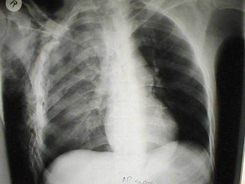
Chest X-ray reveals a pneumatocele as a small, round air-filled area. Computed tomography provides more detailed imaging. Differential diagnoses include lung cancer, tuberculosis, lung abscess in hyper IgE syndrome, a complication of COVID-19 pneumonitis, or an abscess from Staphylococcus aureus infection in cystic fibrosis.
More in Chest Trauma
Sources
Compiled from Wikipedia and the sources listed below. Text from Wikipedia is available under CC BY-SA 4.0; this entry is adapted from it.
- Wikipedia: Pneumatocele (CC BY-SA 4.0).
Spotted an error? Know more?
Reader corrections go straight into our review queue. Suggest an edit · How this site is sourced