Plastid
Membrane-bound organelles derived from endosymbiotic cyanobacteria.
A plastid is a type of membrane-bound organelle found inside the cells of plants, algae, and certain other eukaryotes. Scientists view plastids as endosymbiotic cyanobacteria that now live permanently inside host cells. These organelles are where autotrophic eukaryotes manufacture and store pigments and other key chemical compounds.
The first to name, describe, and clearly define plastids was Andreas Schimper. Each plastid carries its own double-stranded DNA molecule, long thought to be circular like a prokaryotic chromosome, though recent evidence suggests it may sometimes be linear. Plastids produce multiple copies of their own unique genome, called a plastome. The number of genome copies per plastid varies: rapidly dividing new cells may have only a few plastids but over 1,000 genome copies each, while mature cells can contain many plastids with fewer than 100 copies each. A plastome typically encodes transfer RNAs, ribosomal RNAs, and proteins involved in photosynthesis and plastid gene expression. However, these represent only a small fraction of the total proteins needed to build and maintain a plastid; the vast majority are encoded by nuclear genes, and the expression of nuclear and plastid genes is co-regulated.
Plastid DNA exists as protein-DNA complexes called plastid nucleoids, which are localized regions attached to the inner envelope membrane. Unlike a eukaryotic nucleus, a plastid nucleoid lacks a nuclear membrane. Each nucleoid region may contain more than 10 copies of the plastid DNA. In an undifferentiated proplastid, a single nucleoid sits near the center; as the plastid develops or differentiates, many nucleoids appear at the periphery, bound to the inner membrane. During differentiation from one plastid type to another, nucleoids change in shape, size, and location.
Examples of plastids include chloroplasts (used for photosynthesis), chromoplasts (for synthesizing and storing pigments), leucoplasts (non-pigmented plastids that can differentiate further), and apicoplasts (non-photosynthetic plastids found in apicomplexans, derived from secondary endosymbiosis). A permanent primary endosymbiosis event occurred about 1.5 billion years ago in the Archaeplastida clade—which includes land plants, red algae, green algae, and glaucophytes—likely with a cyanobiont related to the genus *Gloeomargarita*. Another primary endosymbiosis happened later, between 140 and 90 million years ago, in the photosynthetic plastids of *Paulinella* amoeboids, involving cyanobacteria from the genera *Prochlorococcus* and *Synechococcus* (the "PS-clade"). Secondary and tertiary endosymbiosis events have also occurred widely, and some organisms have evolved the ability to sequester ingested plastids, a process called kleptoplasty.
In land plants, all plastids derive from proplastids (also called proplasts), which are found in meristematic regions. Proplastids and young chloroplasts typically divide by binary fission, but more mature chloroplasts can also divide. Undifferentiated proplastids can develop into several forms depending on their function: chloroplasts (green, photosynthetic), etioplasts (precursors of chloroplasts), chromoplasts (colored, pigment-storing), gerontoplasts (control dismantling of photosynthetic apparatus during senescence), and leucoplasts (colorless, synthesize monoterpenes). Leucoplasts can further specialize into aleuroplasts, amyloplasts (store starch and detect gravity for geotropism), elaioplasts (store fats), proteinoplasts (store and modify protein), or tannosomes (synthesize tannins and polyphenols). Plastids can differentiate or redifferentiate between these forms depending on morphology and target function.
Many plastids, especially photosynthetic ones, contain numerous internal membrane layers. Chloroplasts perform photosynthesis, converting sunlight into chemical energy while capturing carbon dioxide and releasing oxygen. Other plastids synthesize fatty acids and terpenes, which can be used for energy or as raw materials. For example, plastids in epidermal cells manufacture components of the plant cuticle, including epicuticular wax, from palmitic acid—which itself is synthesized in chloroplasts of the mesophyll. Plastids also store starches, fats, and proteins. In organisms that have lost photosynthetic ability, plastids remain highly useful for manufacturing molecules like isoprenoids.
- field
- Cell biology, endosymbiosis
- known_for
- Photosynthesis, pigment storage, and plastid genome (plastome)
- first_described_by
- Andreas Schimper
Lore & Background
Plastids are membrane-bound organelles that originated from endosymbiotic cyanobacteria, a concept first clearly defined and named by Andreas Schimper. They possess their own double-stranded DNA, long thought to be circular like that of prokaryotes. These organelles are sites for manufacturing and storing pigments and other important chemical compounds, such as isoprenoids, even in organisms that have lost photosynthetic abilities. In land plants, all plastids derive from undifferentiated proplastids located in meristematic regions. Proplastids can differentiate into several forms: chloroplasts, which are green and perform photosynthesis; etioplasts, which are precursors of chloroplasts; chromoplasts, which synthesize and store pigments; gerontoplasts, which dismantle the photosynthetic apparatus during senescence; and leucoplasts, which are colorless and synthesize monoterpenes. Leucoplasts can further specialize into amyloplasts (storing starch and aiding gravity detection), elaioplasts (storing fats), proteinoplasts (storing and modifying protein), aleuroplasts, and tannosomes (synthesizing tannins and polyphenols). Each plastid contains multiple copies of its own genome, or plastome, which encodes transfer RNAs, ribosomal RNAs, and proteins for photosynthesis and gene transcription. However, nuclear genes encode the vast majority of plastid proteins, and their expression is co-regulated with plastid genes. Plastid DNA exists as protein-DNA complexes called nucleoids, which are not surrounded by a membrane. Proplastids have a single central nucleoid, while developing plastids have many nucleoids bound to the inner envelope membrane.
Reader's Guide
Plastids are fundamental to autotrophic eukaryotes, enabling photosynthesis and the synthesis of pigments, fatty acids, terpenes, and other molecules. Their origin as endosymbiotic cyanobacteria is supported by their double-stranded DNA, double membranes, and independent division. The primary endosymbiosis event in Archaeplastida about 1.5 billion years ago gave rise to land plants, red algae, green algae, and glaucophytes. Secondary and tertiary endosymbioses have occurred widely, and some organisms engage in kleptoplasty. Plastids in non-photosynthetic organisms, such as apicoplasts in Apicomplexa, retain biosynthetic functions. The plastid genome may be lost in some parasitic plants, though this is disputed.
Did You Know?
- Plastids are considered intracellular endosymbiotic cyanobacteria.
- A permanent primary endosymbiosis event occurred about 1.5 billion years ago in the Archaeplastida clade.
- Plastid DNA exists as protein-DNA complexes called plastid nucleoids, not surrounded by a nuclear membrane.
The Chemical Architecture of Strand Orientation
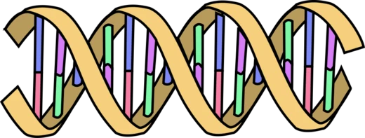
Every single strand of nucleic acid possesses an inherent chemical polarity defined by the numbering of carbon atoms within its pentose sugar ring. At one terminus sits the fifth carbon, which typically bears a phosphate group; at the opposite terminus rests the third carbon, carrying an unmodified hydroxyl substituent. This asymmetry is not merely a naming convention—it is the structural foundation upon which all downstream molecular biology depends. When two strands come together to form a double helix, they must run in opposite orientations. This antiparallel arrangement is what allows complementary bases to pair correctly, and without it, neither replication nor transcription of genetic information would be possible. The convention of labeling these ends as five-prime and three-prime stems directly from the carbon numbering in the ribose or deoxyribose ring, giving molecular biologists a universal language for describing where any given sequence sits along a strand. Whether discussing the location of a gene, a protein-binding site, or a regulatory element, the five-prime-to-three-prime framework provides the coordinate system for all positional language in nucleic acid biology.
The Unidirectional Rule of Polymerization
In living cells, the construction of new nucleic acid strands follows a single, non-negotiable direction: from the five-prime end toward the three-prime end. This constraint arises from the biochemistry of the polymerase enzymes that carry out the task. These enzymes harness the energy released when nucleoside triphosphate bonds are broken, and they use that energy to forge a phosphodiester bond between the incoming nucleoside monophosphate and the three-prime hydroxyl group already present on the growing chain. No known in-vivo mechanism reverses this order. This directional constraint shapes how scientists describe the layout of genetic information. Elements positioned toward the five-prime end are called upstream; those toward the three-prime end are downstream. It also means that directionality and the concept of sense are related but distinct ideas. When a ribosome reads messenger RNA to build a protein, it scans from the five-prime end, and the resulting polypeptide chain grows from its N-terminus toward its C-terminus. In bacteria, mitochondria, and plastids, the first amino acid incorporated is N-formylmethionine rather than plain methionine, a small but notable variation in an otherwise universal reading process.
The Five-Prime End: Capping, Ligation, and Regulatory Architecture
The five-prime terminus of a nucleic acid strand is the carbon-five position of the terminal sugar ring, and it carries outsized functional importance. When a phosphate group is attached there, it becomes the chemical handle that allows two nucleotides to be covalently joined—a process called ligation, in which the five-prime phosphate links to the three-prime hydroxyl of a neighboring nucleotide to create a phosphodiester bond. Molecular biologists exploit this chemistry: by stripping the five-prime phosphate with a phosphatase enzyme, they can prevent a plasmid vector from self-ligating during cloning experiments. In eukaryotic gene expression, the five-prime end of newly made messenger RNA receives a protective cap—a methylated guanosine nucleotide attached through an unusual five-prime-to-five-prime triphosphate linkage. This cap shields the transcript from exonucleases and stabilizes it during translation. The five-prime flanking region upstream of the coding sequence houses the promoter and often enhancers, while the five-prime untranslated region, stretching from the cap site to just before the AUG start codon, contains the ribosome binding site and Kozak sequence, both of which govern how efficiently translation initiates.
The Three-Prime End: Termination, Polyadenylation, and the Sanger Legacy
The three-prime terminus, named for the hydroxyl group on the third carbon of the terminal sugar ring, serves as the active growth point for every new nucleic acid strand. Because polymerases require that free three-prime hydroxyl to attach the next incoming nucleotide, removing it effectively halts chain elongation. Molecular biologists turned this vulnerability into one of the most important tools in genetics: the Sanger dideoxy chain-termination method. By introducing dideoxyribonucleotides that lack the three-prime hydroxyl, researchers can deliberately stop replication at specific points, revealing the precise order of bases in a DNA molecule. At the three-prime end of mature messenger RNA, a tail of fifty to two hundred and fifty adenosine residues is added through polyadenylation. This poly-A tail acts as a timer, influencing how long the transcript survives in the cell and consequently how much protein it ultimately yields. The three-prime untranslated region, spanning from the stop codon to the poly-A tail, carries no protein-coding information yet can modulate both translation efficiency and mRNA stability. The three-prime flanking DNA, once assumed to be silent, is now known to be transcribed and then trimmed away during processing of the primary transcript.
Gallery


Frequently Asked Questions
What is a Plastid?
A plastid is a membrane-enclosed organelle that lives inside the cells of plants, algae, and a few other eukaryotic organisms. It acts as the cell's dedicated workshop and storage depot for pigments and other essential chemical compounds.
What are Plastid's powers or role?
Plastids drive photosynthesis, produce and store pigments, and house the plastome—the organelle's own small genome. Together these functions let autotrophic eukaryotes build their own energy-rich molecules from light and carbon dioxide.
Where did Plastid come from?
Plastids trace their origin to free-living cyanobacteria that were engulfed by an ancestral eukaryotic cell in a single endosymbiotic event. Over billions of years those bacteria lost independence and became permanently embedded as intracellular organelles.
Who first described Plastid?
The Austrian botanist Andreas Schimper is credited with first formally describing plastids. His early observations laid the conceptual groundwork that later cell biologists built on to understand these organelles.
Why is Plastid important in cell & molecular biology?
Plastids are central to the field because they power photosynthesis, sustain most terrestrial food webs, and carry their own genome (the plastome), making them a premier model for studying endosymbiosis, organelle genetics, and pigment biochemistry.
More in Cell And Molecular Biology 1-24
Spotted an error? Know more?
This is a living reference — every entry is fact-audited, and reader corrections feed straight into our audit queue. Suggest an edit · See this site's audit record