Agamid adenovirus
Adenovirus affecting captive bearded dragons, linked to juvenile mortality.
Agamid adenovirus 1, or bearded dragon adenovirus 1, belongs to the Adenoviridae family. It is frequently detected alongside other infections and is linked to higher death rates in both juvenile and adult bearded dragons.
Quick Facts
- Family
- Adenoviridae
- Common name
- Bearded dragon adenovirus 1
- Primary host
- Pogona vitticeps (central bearded dragon)
Facts from the source article.
Infection and consequences
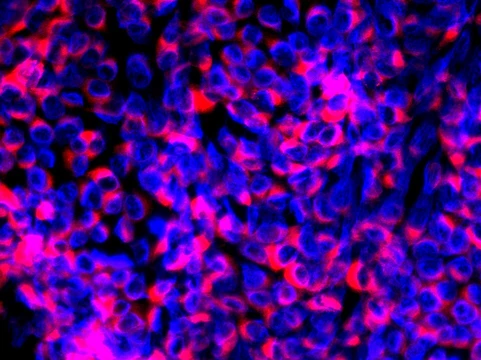
Intranuclear inclusions have been found in the intestinal mucosa, hepatocytes, and bile ducts of infected bearded dragons. Many infected animals show no signs of illness and are considered sub-clinically infected carriers capable of transmitting the virus to others. The virus is known to spread through fecal-oral contact, but additional routes of transmission are suspected based on current research. Agamid adenovirus is becoming widespread in the United States, with several breeders acknowledging infections and ceasing their breeding projects, though some remain unconvinced of its dangers and may sell offspring without testing.
A report from Louisiana State University indicates that baby and juvenile bearded dragons experience high mortality associated with this virus. Suspected or confirmed cases should be isolated and never bred; all animals in contact should also be isolated and not sold. One incident involved a couple handling a breeder's babies while he examined their ill animal, leading to illness and death in his entire clutch ten days later. Another breeder returned from a show and had dying babies test positive within two weeks; subsequently, two other breeders attending the same show lost their colonies.
Sibling clutches have tested both positive and negative, suggesting that some individuals may become infected when eggs pass through the cloaca and pick up viral particles, while others are spared before shelling.
Symptoms
Symptoms of Agamid adenovirus 1 in bearded dragons range from asymptomatic infection to enteritis and death. The dose of infection likely influences clinical disease. Coinfections with agents such as Isospora amphibolouri (a coccidia) and dependovirus (a parvovirus genus) may affect clinical presentation. Husbandry factors including temperature range, diet, population density, and other stressors also likely play significant roles in how the disease manifests.
More in Animal Viral Diseases
Spotted an error? Know more?
Reader corrections go straight into our review queue. Suggest an edit · How this site is sourced