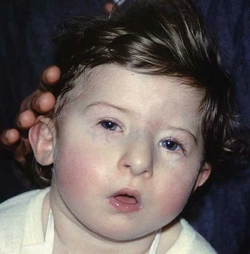
Ectodermal dysplasia
Genetic disorders affecting ectodermal structures like hair, teeth, nails, and glands.
Ectodermal dysplasia (ED) encompasses a group of over 200 genetic disorders marked by abnormal development of structures originating from the embryonic ectoderm, including hair, teeth, nails, and sweat glands. The most common form, hypohidrotic ectodermal dysplasia (HED), affects roughly 1 in 5,000 to 10,000 live births and primarily impacts males due to X-linked inheritance. Mutations in genes such as EDA, EDAR, and EDARADD disrupt the EDA/NF-kappaB signalling pathway, leading to the characteristic symptoms.
Quick Facts
- Most common type
- hypohidrotic ectodermal dysplasia (HED)
- Incidence of hed
- 1 in every 5,000 to 10,000 live births
- Key genes involved
- EDA
- EDAR
- EDARADD
Facts from the source article.
Presentation
Individuals with ectodermal dysplasia often have sparse or absent hair (hypotrichosis), with scalp hair that is fine, brittle, and may lack pigmentation. Eyebrows and eyelashes can also be sparse or missing. Nail abnormalities are common, including thin, brittle, or ridged nails that may split or break; in severe cases nails may be absent or show spoon-like or thickened appearances. The skin is frequently dry and thin, with hyperkeratosis on the palms and soles, and periorbital hyperpigmentation is often present. A major feature in many types is reduced or absent sweat gland function (hypohidrosis or anhidrosis), impairing thermoregulation and risking overheating, especially in hypohidrotic ectodermal dysplasia. Salivary gland underdevelopment (salivary hypoplasia) leads to dry mouth (xerostomia), increasing dental decay risk and complicating swallowing and speech. Dental findings include missing teeth (as seen in orthopantomograms showing absence of ten primary and eleven permanent teeth) and conical-shaped upper incisors that may be restored with composite material.
Diagnosis
Diagnosis is typically based on episodes of unusual fevers from absent or immature sweat glands, along with hair abnormalities and dental findings such as missing or malformed teeth. Clinical diagnosis in early infancy can be difficult because these signs are subtle. Laboratory tests may include quantitative immunoglobulin levels and T-cell subset analysis, as some ED cases with immunodeficiency show hypogammaglobulinemia and impaired cell-mediated immunity. Sweat pore count uses yellow starch-iodine powder on palmar or dorsal skin, turning deep purple where sweating occurs. Skin biopsy can document hypohidrosis and reduced eccrine sweat glands. Molecular genetic testing involves sequencing the EDA gene for X-linked hypohidrotic ED, identifying about 95% of mutations; sequencing EDAR and EDARADD helps detect autosomal dominant and recessive forms. Sequence analysis of EDA cannot detect exonic, multi-exonic, or whole gene deletions in female patients.
Treatment
Treatment focuses on symptom management through a multidisciplinary approach involving dermatologists, dentists, and otolaryngologists. Dental management is central, using prostheses such as dentures, partial dentures, or implants to address missing or malformed teeth. Prosthetic treatment may begin in early childhood to support speech and nutrition, with adjustments as the child grows. Orthodontic interventions manage spacing issues, and maxillofacial surgery may address jaw discrepancies. For young patients, removable prosthetics with expansion screws promote maxillomandibular growth; later, osseointegrated implants offer a more permanent solution if bone development is sufficient. Skin management includes moisturizing creams and emollients to alleviate dryness and hyperkeratosis on palms and soles. Nail care involves trimming and filing to reduce discomfort and damage. Specialized dermatologic treatments may be used for pronounced hyperkeratosis or periorbital pigmentation.
More in Syndromes Affecting Teeth
Spotted an error? Know more?
Reader corrections go straight into our review queue. Suggest an edit · How this site is sourced