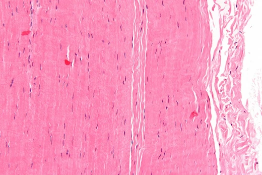
Inflammatory myofibroblastic tumour
Rare intermediate neoplasm of myofibroblastic cells with ALK rearrangements.
Last updated
Inflammatory myofibroblastic tumor (IMT) is a rare neoplasm of mesodermal connective tissue cells. Formerly termed inflammatory pseudotumor, it is now classified by the World Health Organization as an intermediate (rarely metastasizing) fibroblastic and myofibroblastic tumor. IMT is notable for its potential to recur locally, metastasize in a small percentage of cases, and harbor specific genetic alterations.
Quick Facts
- Age peaks (seer study)
- 0–4, 36–40, and >50 years
- Commonest sites (seer study)
- lower limb and hip (22%), upper limb and shoulder (12%), head/face/neck (9%)
- Tumor size range
- 1–25 cm (average 6.5 cm / 2.56 in)
- Metastasis rate
- up to 5% of all cases; up to 10% in ALK fusion-positive cases
Facts from the source article.
Signs and symptoms
IMT can occur in individuals of almost any age and in almost any organ or tissue site. Earlier views that it primarily affected children or young adults and presented in the lung, mesentery, or omentum have been broadened by more recent studies. A retrospective analysis of 92 patients from the SEER program found a mean age of onset of 47.4 years, with peak occurrences at 0–4, 36–40, and over 50 years; middle-aged individuals (41–64 years) constituted one-third of cases.
In that study, the most common sites were the lower limb and hip (22%), upper limb and shoulder (12%), and head, face, and neck (9%). Another study of 25 patients reported the abdomen (40%) and lung/thoracic wall (32%) as the most frequent locations. Individual cases have also been described in the urinary bladder, anal canal, and parameningeal spaces. IMT most often presents as a localized tumor, but distal metastases occur in up to 5% of all cases, or up to 10% of those with ALK fusion protein expression.
Tumor size ranges from 1 to 25 cm (average 6.5 cm), with two-thirds measuring 1.5–6.5 cm. Rare spontaneous regression has been reported.
Molecular abnormalities
In 50–60% of IMT cases and all cases of the epithelioid inflammatory myofibroblastic sarcoma (EIMS) subtype, the neoplastic cells express an abnormal ALK fusion protein resulting from a somatic recombination of the ALK gene. ALK (anaplastic lymphoma kinase) is a serine/threonine-specific protein kinase that activates signaling pathways such as PI3K/AKT/mTOR, Ras GTPase, ERKs, Janus kinase, and STAT proteins, promoting cell growth, proliferation, and survival.
For example, the ALK gene at 2p23 may fuse with the CLTC gene at 17q23 to form a t(2;17)(p23;q23) chimeric gene that produces a constitutively active CLTC-ALK fusion protein. Other genes that fuse with ALK in IMT include TFG, DCTN1, EML4, TPM3, TPM4, ATIC, RANBP2 (most RANBP2-ALK fusions occur in EIMS), CARS1, and SEC31L1. Additionally, about 10% of IMT cases harbor chimeric genes involving ROS1, PDGFRB, or NTRK, whose fusion proteins are also overproduced and overactive, potentially contributing to tumor development.
Treatment
For localized IMT, total surgical resection is commonly recommended, and localized recurrences may also be managed with complete resection. There is little support for adding radiation or systemic chemotherapy to this regimen. Tumors that are unresectable, in inaccessible sites, multifocal, or metastatic are treated with aggressive regimens. In a retrospective study of 59 patients under 25 years old treated with surgery, 31 had no residual disease post-surgery (4 local relapses, mostly re-resected), 19 had microscopic residual disease (treated with various regimens including corticosteroids, vinblastine+methotrexate, ALK inhibitors, or other chemotherapies), and 9 had macroscopic residual disease (treated with vincristine+methotrexate, ALK inhibitors, or other agents).
Among those with microscopic residual disease, 4 had complete responses, 8 partial, 5 stable, and 2 progressive disease; among those with macroscopic residual, none had complete responses, 6 had partial, 1 stable, and 2 progressive. No deaths occurred. Another study of 17 patients (median age 32) with advanced disease treated with Adriamycin-based regimens reported no complete responses, 8 partial, 4 stable, and 5 progressive disease; progression-free survival was 6.6 months and overall survival 21.2 months.
A separate group of 9 patients (median age 16) treated with methotrexate+vinblastine, methotrexate+vinorelbine, or vinblastine+vinorelbine had 2 complete responses, 3 partial, 2 stable, and 2 progressive disease; progression-free survival was not reached and overall survival was 83.4 months.
More in Soft Tissue Disorders
Sources
Compiled from Wikipedia and the sources listed below. Text from Wikipedia is available under CC BY-SA 4.0; this entry is adapted from it.
- Wikipedia: Inflammatory myofibroblastic tumour (CC BY-SA 4.0).
- Word definitions: the , each quoted from its Wikipedia article.
Spotted an error? Know more?
Reader corrections go straight into our review queue. Suggest an edit · How this site is sourced