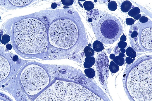
Hereditary sensory and autonomic neuropathy type I
Autosomal dominant neuropathy causing painless foot injuries and ulcerations.
Last updated
Hereditary sensory and autonomic neuropathy type I (HSAN I) is a group of autosomal dominant inherited neurological diseases affecting the peripheral nervous system, particularly sensory and autonomic functions. Its hallmark is marked loss of pain and temperature sensation in the distal lower limbs, often leading to unrecognized injuries and foot ulcerations. It is the most common type among the five types of HSAN.
Quick Facts
- Inheritance
- Autosomal dominant
- Age of onset
- Between adolescence and adulthood (2nd to 5th decade); congenital or childhood onset occasionally reported
- Subtypes
- HSAN IA–E
- Associated gene (hsan ia)
- SPTLC1
- Enzyme encoded
- Serine palmitoyltransferase (SPT)
Facts from the source article.
History
The first descriptions of sporadic and familial cases compatible with HSAN appeared in 19th-century French literature, with the familial case featuring ulcers on the soles of the feet. In 1922, Hicks described a similar condition in a London family where 10 persons had perforating foot ulcers, lancinating and shooting pains, and deafness. Later, Jughenn et al. and Denny-Brown demonstrated that the underlying pathology was a neuropathy, not an anatomical disorder.
Many other familial conditions with similar features were subsequently reported. Early names included mal perforant du pied, ulcero-mutilating neuropathy, hereditary perforating ulcers, familial trophoneurosis, familial syringomyelia, and hereditary sensory radicular neuropathy. In dermatological literature, Thèvenard syndrome is used for familial forms, and Bureau-Barrière syndrome for sporadic forms. In 1975, Dyck and Otha proposed a descriptive classification, introducing the term hereditary sensory neuropathy (HSN), later changed to hereditary sensory and autonomic neuropathy (HSAN) due to substantial autonomic involvement.
They categorized the diseases into five types (HSAN I–V) based on inheritance, clinical features, and age at onset; HSAN I comprises those with autosomal dominant inheritance and adolescence or adulthood onset.
Signs and symptoms
Affected individuals often first notice loss of pain and temperature sensation, or all sensory modalities, in the feet. As the disease progresses, sensory abnormalities may extend up to the knees, though many do not recognize the loss for a long time. Awareness typically comes from painless injuries, burns, or slowly healing wounds and foot ulcers. Ulcerations may arise from permanent pressure, such as long walks or poorly fitting shoes.
Minor wounds or blisters can develop into deep foot ulcerations; once infection occurs, complications like inflammation and destruction of underlying bones may follow. Some individuals who do not lose sensation experience spontaneous pain, and many exhibit variable degrees of symmetrical distal muscle weakness and wasting. Sensory disturbances primarily involve loss of pain and temperature sensation in distal lower limbs, which can extend proximally and to upper limbs over time.
Others instead suffer severe shooting, burning, or lancinating pains in limbs or trunk. Autonomic disturbances, if present, manifest as decreased sweating. Motor disturbances range from absent to severe distal muscle weakness and wasting, even within families. The disease progresses slowly but often severely disables individuals after a long duration.
Minor injuries in painless areas can result in slow-healing wounds that become chronic ulcerations. Infection can lead to foot deformity, inflammation of bones, spontaneous fractures, and progressive joint degeneration. Foot deformity promotes hyperkeratosis at pressure points, and complications may necessitate amputation.
Cause
Advances in molecular genetics have identified most genes associated with HSAN I, but the underlying molecular mechanisms are not fully understood. A challenge is elucidating how faulty genes involved in basic cellular functions—such as sphingolipid metabolism, organellar integrity, membrane dynamics, and transcription regulation—affect specific neuronal populations. HSAN IA is associated with heterozygous missense mutations in the SPTLC1 gene, which encodes SPTLC1 protein.
Together with SPTLC2, this forms serine palmitoyltransferase (SPT), a pyridoxal-5'-phosphate-dependent enzyme catalyzing the first and rate-limiting step in de novo sphingolipid biosynthesis. Sphingolipids, with cholesterol, form lipid rafts in plasma membranes that facilitate signal transduction. Initially, SPTLC1 mutations were thought to increase SPT activity, but later studies suggested reduced activity. However, reduced activity cannot account for aberrant sphingolipid features in heterozygous patient-derived cells or clinical features in heterozygous mice, indicating that non-mutant SPTLC1 may suffice for normal biosynthesis.
Neuronal degeneration likely results from subtle long-term effects of mutations or accumulation of toxic lipids produced by mutant enzymes.
Diagnosis
Diagnosis of HSAN I is based on symptom observation and supported by a family history suggesting autosomal dominant inheritance. Additional tests include nerve conduction studies in the lower limbs to confirm sensory and motor neuropathy. In sporadic cases, acquired neuropathies such as diabetic foot syndrome and alcoholic neuropathy can be excluded using magnetic resonance imaging and interdisciplinary discussion among neurologists, dermatologists, and orthopedists.
Identification of causative genes has revolutionized diagnosis, which now relies on detecting mutations by direct sequencing of the genes. Accurate phenotyping of patients remains crucial. For pregnant patients, termination of pregnancy is not recommended. HSAN I must be distinguished from hereditary motor and sensory neuropathy (HMSN) and other HSAN types (II–V).
Prominent sensory abnormalities and foot ulcerations separate HSAN I from HMSN. HSAN II differs by autosomal recessive inheritance, earlier onset, diffuse sensory loss to the whole body, and fewer or no motor symptoms. HSAN III–V are distinguished by congenital onset and typical features such as predominant autonomic disturbances in HSAN III or congenital loss of pain and anhidrosis in HSAN IV. In 1993, Peter James Dyck divided HSAN I into five subtypes (IA–E) based on additional features, thought to arise from genetic diversity or environmental factors; molecular genetic studies later confirmed this diversity.
Did You Know?
- Gene-based therapies are not available; supportive care is the only treatment.
- Treatment for foot ulcers and infections can follow guidelines for diabetic foot care.
- The disease progresses slowly and does not influence life expectancy if signs and symptoms are properly treated.
More in Peripheral Nervous System Disorders
Sources
Compiled from Wikipedia and the sources listed below. Text from Wikipedia is available under CC BY-SA 4.0; this entry is adapted from it.
- Wikipedia: Hereditary sensory and autonomic neuropathy type I (CC BY-SA 4.0).
- Word definitions: the , each quoted from its Wikipedia article.
Spotted an error? Know more?
Reader corrections go straight into our review queue. Suggest an edit · How this site is sourced