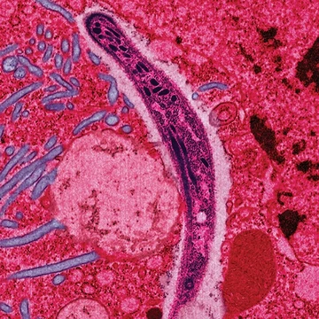
Lymphedema
Localized swelling from lymphatic system compromise, often from cancer or parasites.
Lymphedema is a condition of localized swelling caused by a compromised lymphatic system, which is critical for immune function and returning interstitial fluid to the bloodstream. It is most frequently a complication of cancer treatment or parasitic infections, though it can also arise from genetic disorders. Tissues affected by lymphedema are at high risk of infection due to the impaired lymphatic system.
Quick Facts
- Field
- Vascular medicine
- Rheumatology
- Physical medicine and rehabilitation
- General surgery
- Plastic surgery
- Diagnosis
- Based on symptoms
- Differential
- Lipodystrophy, venous insufficiency
Facts from the source article.
Causes and risk factors
Lymphedema may be inherited (primary) or caused by injury to lymphatic vessels (secondary). Risk factors include old age, being overweight or obese, and having rheumatic or psoriatic arthritis. A body mass index above 30 increases the risk nearly threefold when radiation therapy accompanies breast-conserving mastectomy. Secondary lymphedema most commonly follows lymph node dissection, surgery, or radiation therapy for cancer, especially breast cancer; radiation's impact on lymphatic vessels appears later as fibrotic scarring restricts flow. Damage from infections such as cellulitis or, in tropical areas, parasitic filarial worms (filariasis) can also cause secondary lymphedema. Primary lymphedema may be congenital or sporadic, associated with syndromes like Turner syndrome, Milroy's disease, or Klippel–Trénaunay syndrome, often due to absent or malformed lymph nodes or channels. It can appear at birth, at puberty (praecox), or later in adulthood (tarda). In men, lower-limb primary lymphedema is most common.
Physiology
Lymph originates from fluid filtering out of blood, containing proteins, cellular debris, and bacteria. Blind-ended endothelial-lined vessels with fenestrated openings collect this fluid, allowing entry of particles as large as cells. Inside the lumen, fluid moves through increasingly larger vessels; early vessels have rudimentary valves, later ones have complete valves similar to venous valves. Once in fully valved vessels, rhythmic peristaltic contractions of smooth muscle cells in the vessel walls pump lymph, with the sympathetic nervous system regulating contraction frequency and power. Passive forces such as nearby muscle contraction, arterial pulse pressure, and the chest cavity's respiratory vacuum contribute only a minor percentage of lymph transport. Collected fluid passes through lymph nodes, which remove debris and monitor for microbes, then drains into the thoracic duct or right lymphatic duct and into the blood circulation. Chronic inflammation is hypothesized to be a key regulator in lymphedema development, with Th2 differentiation playing a crucial role. Increased expression of Th2-inducing cytokines in epidermal cells of lymphoedematous limbs has been observed. Treatment with QBX258 reduced hyperkeratosis and fibrosis, decreased CD4+ cell numbers, and normalized Th2-inducing cytokine and IL13R expression by keratinocytes, suggesting epidermal cells may initiate or coordinate chronic Th2 responses. Sustained lymphatic stasis leads to CD4+ T-cell infiltration, causing inflammation and fibrosis.
Diagnosis
Diagnosing lymphedema starts with evaluating symptoms and signs, and tests are mainly used to exclude other possibilities. Getting the stage and diagnosis right can help with treatment planning. Swelling in a limb can come from many different issues, each needing its own approach. Right now, diagnosis depends on the patient’s history, a physical exam, and measuring the limb. Imaging like lymphoscintigraphy or indocyanine green lymphography is only used if surgery might be needed. There’s debate over the best staging method because several systems exist. The exam begins by looking at the limb: checking color, hair growth, visible veins, size, and any sores or ulcers. Missing hair might point to arterial problems. The limb’s circumference is tracked over time. In early stages, raising the limb can sometimes ease swelling. Pressing on the wrist or ankle helps gauge the swelling, and pulses are felt. Lymph nodes in the armpit or groin may be enlarged. If they stay swollen for more than three weeks, it could signal an infection or another issue, such as a complication from breast cancer surgery, and needs further checking.
Treatment
Curative treatment for lymphedema is surgical, involving supermicrosurgery such as lymph node transplant from a healthy donor site to the diseased area. In simpler cases, a lymphatic-venous shunt drains excess lymph into adjacent superficial veins. These microsurgical procedures result in improvement in 98% of cases. Complementary treatment may improve outcomes and commonly includes compression therapy, good skin care, exercise, manual lymphatic drainage (MLD), and use of an intermittent pneumatic compression pump, collectively known as combined decongestive therapy. MLD is most effective in mild to moderate disease; in breast cancer-related lymphedema, it is safe and may add benefit to compression bandages. Most people can be managed conservatively. Diuretics are not useful. Surgery is generally reserved for cases not improved by other measures. Compression is imperative after diagnosis; elastic compression garments are worn all day but may be removed for sleep unless otherwise prescribed. Inelastic garments provide containment and reduction. Compression bandaging uses layers of padding and short-stretch bandages, as long-stretch bandages cannot produce proper therapeutic tension and may cause a tourniquet effect. Compression bandages provide resistance that assists pumping fluid out during exercise, increasing lymphatic drainage and reducing swelling.
More in Parasitic Diseases
Spotted an error? Know more?
Reader corrections go straight into our review queue. Suggest an edit · How this site is sourced