Excitotoxicity
Neuronal injury from excessive glutamate receptor stimulation.
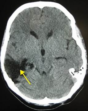
Excitotoxicity is a pathological process in which nerve cells are damaged or killed by excessive stimulation of neurotransmitter receptors, most notably by high levels of glutamate. This overactivation allows large amounts of calcium ions to enter the cell, triggering enzymes that degrade cellular structures. It is implicated in a wide range of conditions, including stroke, traumatic brain injury, spinal cord injury, and neurodegenerative diseases.
Quick Facts
- First observed
- 1954 by T. Hayashi
- Term coined
- 1969 by John Olney
- Key receptor types
- NMDA and AMPA
- Primary endogenous excitotoxin
- Glutamate
Facts from the source article.
History
The harmful effects of glutamate on the central nervous system were first observed in 1954 by T. Hayashi, who noted that direct application of glutamate caused seizure activity, though this report went unnoticed for several years. In 1957, D. R. Lucas and J. P. Newhouse observed that a subcutaneous dose described as "a little less than lethal" destroyed neurons in the inner layers of the retina in newborn mice. John Olney in 1969 found that the phenomenon occurred throughout the brain, coined the term excitotoxicity, and determined that cell death was restricted to postsynaptic neurons, that glutamate agonists were as neurotoxic as their efficiency to activate glutamate receptors, and that glutamate antagonists could stop the neurotoxicity. In 2002, Hilmar Bading and co-workers found that excitotoxicity is caused by activation of NMDA receptors located outside synaptic contacts. In 2020, the same group described a death signaling complex consisting of extrasynaptic NMDA receptor and TRPM4; disruption of this complex using NMDAR/TRPM4 interface inhibitors renders extrasynaptic NMDA receptor non-toxic.
Pathophysiology
Excitotoxicity can result from endogenous excitotoxins, with glutamate being a prime example and the major excitatory neurotransmitter in the mammalian central nervous system. Under normal conditions, glutamate concentration in the synaptic cleft can rise to 1 mM but is rapidly decreased within milliseconds. When the concentration cannot be lowered or reaches higher levels, the neuron undergoes apoptosis. This phenomenon can occur after brain injury and spinal cord injury; within minutes after spinal cord injury, damaged neural cells spill glutamate into the extracellular space, where it can stimulate presynaptic receptors to enhance further release. Brain trauma or stroke can cause ischemia, leading to accumulation of glutamate and aspartate in extracellular fluid, causing cell death aggravated by lack of oxygen and glucose. The resulting biochemical cascade is called the ischemic cascade. Increased extracellular glutamate activates Ca2+ permeable NMDA receptors on myelin sheaths and oligodendrocytes, making them susceptible to calcium influx and excitotoxicity. Excess calcium in the cytosol initiates apoptosis through cleaved caspase processing and also opens the mitochondrial permeability transition pore, causing mitochondria to swell and release reactive oxygen species and other proteins that can lead to apoptosis. The pore can also cause mitochondria to release more calcium, and production of adenosine triphosphate may stop, with ATP synthase possibly beginning to hydrolyse ATP instead of producing it.
More in Neurotrauma
Spotted an error? Know more?
Reader corrections go straight into our review queue. Suggest an edit · How this site is sourced