Groin
Junctional area between torso and thigh, site of adductor muscles.

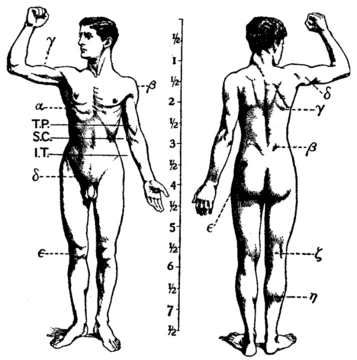
The groin, referred to anatomically as the inguinal or iliac region, is where the torso connects to the thigh. Located on the front of the body, it sits on each side of the pubic tubercle, marking the spot where the lower abdominal wall meets the thigh. This junction creates a visible fold called the inguinal groove or crease. It also corresponds to the medial compartment of the thigh, which holds the attachment points for the hip adductor muscles—often called the groin muscles.
These adductor muscles include the adductor brevis, adductor longus, adductor magnus, gracilis, and pectineus. Their primary action is to adduct the thigh, pulling the thigh and knee toward the body’s midline. Nerve supply to the groin comes from branches of the lumbar plexus. Specifically, the femoral nerve innervates the pectineus, while the tibial nerve supplies the hamstring portion of the adductor magnus.
Beneath the skin in the groin lie three to five deep inguinal lymph nodes, which are part of the immune system. A chain of superficial inguinal lymph nodes drains into these deeper nodes. Within the inguinal triangle, two depressions exist: the lateral inguinal fossa and the medial inguinal fossa. The inguinal ligament, which runs from the pubic tubercle to the anterior superior iliac spine, is a key structure in hernia surgery.
Clinically, a pulled groin muscle typically refers to a painful strain of the hip adductor muscles, often linked to overuse or a prior injury. An inguinal hernia is a hernia in this region, classified as direct or indirect based on its exact location. When both types occur on the same side, it is called a saddlebag hernia. Like other flexion surfaces of large joints—such as the popliteal fossa, armpit, and cubital fossa—the groin is an area where blood vessels and nerves run relatively close to the surface and contain an increased number of lymph nodes. In venography, the groin is the preferred site for making an incision to insert a catheter into the vascular system.
- field
- Human anatomy
- known_for
- Junctional area between torso and thigh; site of groin muscles, inguinal lymph nodes, and inguinal hernias
- location
- Front of body, on either side of pubic tubercle
- components
- Adductor muscles (adductor brevis, adductor longus, adductor magnus, gracilis, pectineus), inguinal ligament, deep and superficial inguinal lymph nodes, lateral and medial inguinal fossae
Lore & Background
The groin, or inguinal region, is the junctional area on the front of the body where the torso meets the thigh, located on either side of the pubic tubercle. A natural fold, known as the inguinal groove or crease, marks this junction. This region encompasses the medial compartment of the thigh, which contains the attachments of the hip adductor muscles—collectively called the groin muscles. These muscles include the adductor brevis, adductor longus, adductor magnus, gracilis, and pectineus, and their primary action is to adduct the thigh, drawing it and the knee toward the body’s midline. Nerve supply to the groin comes from branches of the lumbar plexus; specifically, the femoral nerve innervates the pectineus, while the tibial nerve supplies the hamstring portion of the adductor magnus. Beneath the skin, the groin houses three to five deep inguinal lymph nodes, which are part of the immune system, and a chain of superficial inguinal lymph nodes drains into these deeper nodes. The inguinal triangle contains two depressions called fossae: the lateral inguinal fossa and the medial inguinal fossa. The inguinal ligament, which extends from the pubic tubercle to the anterior superior iliac spine, is a key anatomical landmark, particularly relevant for hernia surgeries. Like other flexion surfaces of large joints, such as the armpit or popliteal fossa, the groin is an area where blood vessels and nerves pass relatively close to the surface and where lymph nodes are abundant. A common injury in this area is a groin pull, a strain of the leg adductor muscles, often resulting from overuse or prior injury. Inguinal hernias are also clinically significant and can be classified as direct or indirect based on their location; a direct and indirect hernia occurring together on the same side is termed a saddlebag hernia. In medical procedures like venography, the groin is the preferred site for catheter insertion into the vascular system.
Reader's Guide
The groin is clinically significant as the site of common injuries and conditions. A pulled groin muscle usually refers to a painful strain of the hip adductor muscles, related to risk factors including overuse and previous injury. Inguinal hernias occur in the groin and can be direct or indirect, sometimes presenting together as a saddlebag hernia. The groin is also an area where blood vessels and nerves pass relatively superficially, with an increased amount of lymph nodes, and in venography it is the preferred site for incisions to enter a catheter into the vascular system. The inguinal ligament, running from the pubic tubercle to the anterior superior iliac spine, is important for hernia operations.
Did You Know?
- The groin contains three to five deep inguinal lymph nodes that play a role in the immune system.
- A 'groin pull' involves a strain of the leg adductor muscle group in the inguinal area.
- The inguinal ligament runs from the pubic tubercle to the anterior superior iliac spine.
- In a venography procedure, the groin is the preferred site for incisions to enter a catheter into the vascular system.
Frequently Asked Questions
What is the Groin in the musculoskeletal system?
The Groin, also called the inguinal or iliac region, is the transitional zone where the lower abdominal wall connects to the upper thigh. It sits at the front of the body on both sides of the pubic tubercle, marked by a visible crease known as the inguinal groove.
Where exactly is the Groin located?
It occupies the junction between the torso and the thigh, positioned on either side of the pubic tubercle at the front of the body. The lower abdominal wall meets the thigh here, creating the characteristic inguinal groove.
Which muscles are part of the Groin region?
The adductor muscle group—comprising the adductor brevis, adductor longus, adductor magnus, gracilis, and pectineus—anchors in this area. The inguinal ligament also forms a key structural component of the region.
Why is the Groin a common site for hernias?
The junction between the abdominal wall and the thigh creates a natural weak point where internal structures can protrude outward. This is why inguinal hernias are among the most frequently encountered types of hernia in clinical practice.
What other important structures reside in the Groin?
Beyond the adductor muscles, the region houses both deep and superficial inguinal lymph nodes, which serve as drainage points for the lower limbs and external genitalia. The lateral and medial inguinal fossae are also anatomical landmarks found here.
More in Musculoskeletal System 1-24
Spotted an error? Know more?
This is a living reference — every entry is fact-audited, and reader corrections feed straight into our audit queue. Suggest an edit · See this site's audit record