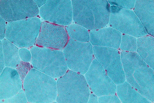
Glutaric aciduria type 1
A neurometabolic disorder causing brain damage and secondary carnitine deficiency.
Glutaric acidemia type 1 (GA1) is an inherited disorder in which the body cannot fully break down the amino acids lysine, hydroxylysine and tryptophan. Accumulation of intermediate breakdown products can damage the brain, particularly the basal ganglia, and causes secondary carnitine deficiency. It is an autosomal recessive condition resulting from deficiency of the enzyme glutaryl-CoA dehydrogenase.
Quick Facts
- Prevalence
- approximately 1 in 30,000 to 40,000 births
- Inheritance
- autosomal recessive
- Gene
- GCDH
- Enzyme
- glutaryl-CoA dehydrogenase
- Higher prevalence populations
- Amish community and Ojibway population of Canada (up to 1 in 300 newborns)
Facts from the source article.
Did You Know?
- Relatives of children with GA1 can have low GCDH activity; in an early study, GCDH activity was found to be 38%, 42%, and 42% in three of the four unaffected relatives tested.
- Normally, magnetic resonance imaging shows the Sylvian fissure to be operculated, but in GA1-associated encephalopathy, operculation is absent.
- A 2006 study of 279 patients found that of those with symptoms (185, 66%), 95% had suffered an encephalopathic crises, usually with following brain damage.
Signs and symptoms
Glutaric aciduria type 1 affects people very differently; some have mild symptoms while others face serious challenges. The condition can be identified in two ways: when it is found before or at birth and managed with diet, or when it is discovered only after a brain crisis. Babies with this disorder are often born with an unusually large head, which is one of the earliest clues. Because of this, any unexplained large head should be checked for a GCDH deficiency to catch the disease early. People with GA1 may have trouble moving, along with spasms, jerking, stiffness, or weak muscle tone, sometimes due to a lack of carnitine. After a crisis, the condition can resemble a genetic form of cerebral palsy. Special wheelchairs are used to manage striatal damage by limiting abnormal movements, though restraint can increase spasticity. Some individuals also experience brain or eye bleeding that might be mistaken for child abuse.
Pathophysiology
Glutaryl-CoA dehydrogenase participates in the degradation of lysine, hydroxylysine and tryptophan. This enzyme deficiency allows glutaric acid, 3-hydroxyglutaric acid and to a lesser extent glutaconic acid to build up to abnormal levels. Glutaric acid can block Na+-dependent glutamate uptake by causing oxidative stress; inhibition of Na+/K+-ATPase hampers astrocyte re-uptake of glutamate and worsens excitotoxicity. Due to metabolic dysfunction, alpha-ketoglutarate gets depleted which hampers HIF1a degradation and upregulates VEGF, causing weak blood vessels that tend to cause hemorrhages. Oxidative stress damages lipids, proteins and DNA. GA also activates astrocytes and can cause microgliosis. 3-Hydroxyglutaric Acid can cause oxidative stress, inhibition of mitochondrial complex II, and astrogliosis, and can inhibit glutamate decarboxylase which participates in synthesis of GABA.
Treatment
Like many other organic acidemias, GA1 causes carnitine depletion. Whole-blood carnitine can be raised by oral supplementation, but this does not significantly change blood concentrations of glutarylcarnitine or esterified carnitine, suggesting that oral supplementation is suboptimal in raising tissue levels. In contrast, regular intravenous infusions of carnitine cause distinct clinical improvements. Choline increases carnitine uptake and retention and can increase exercise tolerance, truncal tone and general well-being. Dietary control may help limit progression of the neurological damage. Lysine restriction, as well as carnitine supplementation, are considered the best predictors of a good prognosis for GA1, excluding patients who already suffered an encephalopathic crisis. Vegetarian diets and breastfeeding are common ways to limit protein intake.
Epidemiology
Glutaric aciduria type 1 is classified in multiple ways: as a metabolic disorder, a neurometabolic disease, a form of cerebral palsy, or a basal ganglia disorder. It can also be mistaken for shaken baby syndrome. Treatment depends on the chosen framework, focusing either on restricting certain precursors or on neurorehabilitation. Acute striatal necrosis, a key pathological feature, appears in over twenty other conditions with widely different causes, including HIV encephalopathy, pneumococcal meningitis, and methylmalonic acidemia. In a reported group of 279 individuals with GA1, 185 showed symptoms, which was considered a sign of poor treatment response. For those without affected relatives or newborn screening, macrocephaly is the primary indicator. Though GA1 is treatable, about two-thirds of patients with GA1 encephalopathy gain little from standard GA1 therapy. However, they may benefit from interventions used for other basal ganglia disorders, such as brain implants, stem cell therapies, growth factors, and monoaminergic agents.
More in Mitochondrial Diseases
Spotted an error? Know more?
Reader corrections go straight into our review queue. Suggest an edit · How this site is sourced