Drug-induced autoimmune hemolytic anemia
A rare immune-mediated anemia triggered by drugs, often requiring prompt recognition and cessation of the offending agent.
Last updated

Spicy · CC BY-SA 4.0
Drug-induced autoimmune hemolytic anemia, or DIIHA, is a rare condition where the immune system attacks the body's own red blood cells after exposure to certain medications. It is hard to tell apart from other anemias, often delaying care. Stopping the responsible drug is the main treatment. Many antibiotics can trigger it, and it occurs in about one to two people per million globally.
Background
In some cases, a drug can cause the immune system to mistakenly think the body's own red blood cells are dangerous, foreign substances. Antibodies then develop against the red blood cells.
The antibodies attach to red blood cells and cause them to break down too early. As of 2020 over 130 drugs have been reported to cause DIIHA. The list includes: Cephalosporins (a class of antibiotics), Dapsone, Levofloxacin, Methyldopa, Nitrofurantoin, Nonsteroidal anti-inflammatory drugs (NSAIDs) - among them, the commonly used Diclofenac and Ibuprofen, Phenazopyridine (pyridium), Quinidine.
The Oxidative Assault on Red Blood Cells
When certain medications enter the bloodstream, they can trigger a cascade of intracellular oxidizing radicals—molecules like hydrogen peroxide—that attack the structural integrity of red blood cells. These radicals interact directly with hemoglobin and the cell membrane, crosslinking proteins and initiating lipid peroxidation that weakens the delicate bilayer. Over time, this oxidative damage promotes the formation of Heinz bodies, abnormal inclusions that further compromise the cell's ability to function.
Unlike the autoimmune variant, where the immune system itself turns against red cells, this nonautoimmune pathway is purely chemical: the drug or its metabolites act as the aggressor. The result is a progressive destruction of red cells that the bone marrow struggles to outpace, particularly when the insult is acute and severe. This mechanism explains why the condition can strike without any prior immune sensitization, making it a distinct clinical entity from antibody-mediated hemolysis.
Genetic Vulnerability and the Antioxidant Shield
Red blood cells depend on robust antioxidant defense systems to neutralize the oxidizing radicals produced during normal metabolism. In individuals carrying a deficiency in glucose-6-phosphate dehydrogenase, the enzyme responsible for generating NADPH—the key antioxidant molecule—operates at reduced capacity, leaving cells far more vulnerable to exogenous oxidative insults. Similarly, those with Hemoglobin H disease, a condition resulting from the loss of three alpha-globin chain genes, produce abnormal hemoglobin that simultaneously increases the generation of oxidizing radicals while impairing the cell's ability to withstand them.
This double hit explains why drugs such as primaquine, dapsone, nitrofurantoin, rasburicase, and pegloticase, which are generally well tolerated in the broader population, can precipitate dramatic hemolytic episodes in these genetically predisposed individuals. The interplay between pharmacologic oxidative stress and inherited antioxidant fragility lies at the heart of this condition's clinical significance.
From First Symptoms to Life-Threatening Crisis
The clinical picture of drug-induced nonautoimmune hemolytic anemia can emerge abruptly after a single drug exposure or gradually over days to weeks, and its severity spans a wide spectrum. Early manifestations mirror those of any acute anemia: pallor, fatigue, dizziness, tachycardia, shortness of breath, and even syncope. As hemolysis intensifies, patients develop the hallmark signs of red cell destruction—abdominal and back pain, jaundice, and urine that turns dark or frankly red from free hemoglobin.
In the most devastating presentations, the process escalates into shock, disseminated intravascular coagulation, acute renal failure, or even death. The rapidity with which symptoms can progress, combined with the fact that initial production of new red cells by the bone marrow cannot keep pace with ongoing destruction, makes early recognition critical. The condition's unpredictability and potential for catastrophic decompensation underscore why it must be distinguished promptly from other causes of hemolytic anemia.
Unmasking the Diagnosis and Breaking the Cycle
Because the onset can be sudden and severe, the initial diagnostic workup centers on a complete blood count paired with a peripheral blood smear. Hemoglobin and hematocrit values typically fall as the marrow's compensatory erythropoiesis lags behind the pace of destruction, while reticulocyte counts rise as the bone marrow ramps up production. Biochemical markers of hemolysis—elevated lactate dehydrogenase, increased unconjugated bilirubin, and depleted haptoglobin—provide corroborating evidence, and a urine dipstick positive for heme further supports ongoing red cell breakdown.
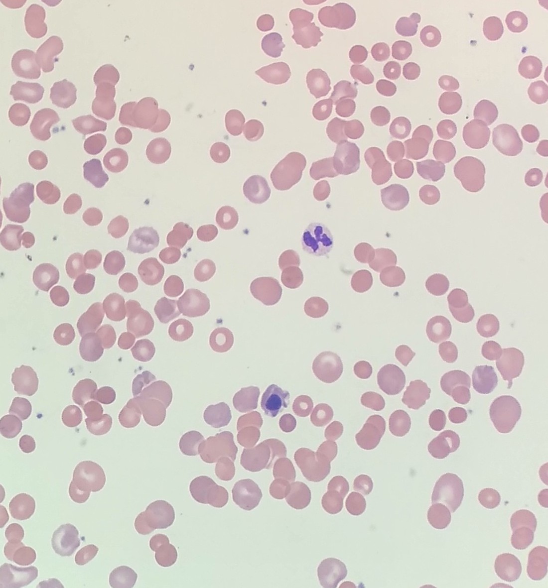
Treatment, once the offending agent is identified, is fundamentally straightforward: discontinue the suspected drug and provide supportive care, including aggressive hydration and, when necessary, red blood cell transfusions to restore oxygen-carrying capacity. In cases complicated by thrombotic microangiopathy, managing the underlying vascular injury becomes an additional priority alongside hemolysis control.
More in Drug-induced diseases
Sources
Compiled from Wikipedia and the sources listed below. Text from Wikipedia is available under CC BY-SA 4.0; this entry is adapted from it.
- Wikipedia: Drug-induced autoimmune hemolytic anemia (CC BY-SA 4.0).
- Word definitions: the , each quoted from its Wikipedia article.
Spotted an error? Know more?
Reader corrections go straight into our review queue. Suggest an edit · How this site is sourced