Adenine phosphoribosyltransferase deficiency
A rare metabolic disorder causing kidney stones and renal failure.
Adenine phosphoribosyltransferase deficiency is a rare autosomal recessive metabolic disorder caused by mutations of the APRT gene. The condition leads to the accumulation of 2,8-dihydroxyadenine, resulting in urolithiasis and renal failure. It is classified into two types, with type two predominantly found in Japan.
Quick Facts
- Complications
- End-stage renal disease.
- Onset
- Infancy to late adulthood.
- Types
- Type 1 and type 2.
- Causes
- Mutations in the APRT gene.
- Diagnosis
- Urine microscopy and kidney stone analysis.
- Differential
- Uric acid nephrolithiasis
- Xanthinuria
- and Primary hyperoxaluria.
- Medication
- Allopurinol.
- Frequency
- 0.4% to 1.2%
Facts from the source article.
Signs and symptoms
Adenine phosphoribosyltransferase deficiency affects only the kidneys and urinary tract, causing stones, blood in the urine, infections, and both acute and chronic kidney disease. It can appear at any age, from infancy to over 70. Some people never develop symptoms and are only found through family screening. In 15% of adult cases, kidney failure leads to dialysis or transplant. Infants may have reddish-brown diaper stains. The condition involves dihydroxyadenine crystals forming in kidney tissue, which can cause severe damage. Dehydration may trigger sudden kidney failure by concentrating the urine and promoting crystal buildup.
Causes
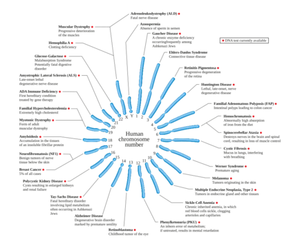
Adenine phosphoribosyltransferase deficiency is an autosomal recessive condition, requiring two copies of the mutated gene. The APRT gene is located on chromosome 16q24, contains five exons, and spans 2.8 kb of DNA with a coding region of 540 bp. Complete deficiency develops in individuals with mutations in both copies of the gene. There is no evidence that genotype correlates with phenotype; environmental factors or modifiers may account for heterogeneity.
Mechanism
All tissues express the APRT enzyme, which is the sole route for recovering adenine from dietary and polyamine biosynthesis sources. In the absence of functional APRT, adenine is converted to 8-hydroxyadenine, which is further metabolized by xanthine dehydrogenase to dihydroxyadenine. Dihydroxyadenine has high renal clearance and may be secreted tubularly. It precipitates in renal parenchyma, forming insoluble crystals that can accumulate and cause crystalline nephropathy.
Diagnosis
Diagnosis is based on identification of dihydroxyadenine by kidney stone analysis or urine crystal examination. Infrared spectroscopy combined with morphologic examination under a stereomicroscope identifies dihydroxyadenine in all cases of kidney stones. Biochemical stone analysis is unreliable and cannot distinguish dihydroxyadenine from uric acid. Light and polarizing microscopy of crystalluria is a noninvasive and inexpensive method. Morning urine samples are ideal for studying crystalluria; crystal counts per volume are higher in untreated patients.
More in Autosomal Recessive Disorders
Spotted an error? Know more?
Reader corrections go straight into our review queue. Suggest an edit · How this site is sourced